Goat anti-Rabbit IgG (Heavy & Light Chain) Antibody (FITC)
 (31 references)
 (1 validation)
-
- Target See all IgG products
- IgG
-
Binding Specificity
- Heavy & Light Chain
-
Reactivity
- Rabbit
-
Host
- Goat
-
Clonality
- Polyclonal
-
Conjugate
- FITC
-
Application
- Western Blotting (WB), Flow Cytometry (FACS), FLISA, Fluorescence Microscopy (FM), Dot Blot (DB)
- Supplier Product No.
- 611-1202
- Supplier
- Rockland
- Purpose
- Rabbit IgG (H&L) Secondary Antibody Fluorescein Conjugated
- Cross-Reactivity (Details)
- Assay by immunoelectrophoresis resulted in a single precipitin arc against anti-Fluorescein, anti-Goat Serum, Rabbit IgG and Rabbit Serum.
- Characteristics
- Anti-Rabbit IgG F(ab')2 Antibody generated in goat recognizes the dimeric Fab portion of the rabbit IgG molecule. Rabbit IgG F(ab')2 is a proteolytic fragment of immunoglobulin G (IgG) obtained by limited digestion with the enzyme pepsin under controlled conditions of temperature, time and pH . F(ab')2 Molecules lack the Fc portion of IgG and therefore receptors that bind rabbit IgG F(c) will not bind rabbit IgG F(ab')2 Molecules. Secondary Antibodies are available in a variety of formats and conjugate types. When choosing a secondary antibody product, consideration must be given to species and immunoglobulin specificity, conjugate type, fragment and chain specificity, level of cross-reactivity, and host-species source and fragment composition.
- Purification
- This product was prepared from monospecific antiserum by immunoaffinity chromatography using Rabbit IgG coupled to agarose beads followed by solid phase adsorption(s) to remove any unwanted reactivities.
- Immunogen
- Optional[Immunogen]: Rabbit IgG whole molecule
- Isotype
- IgG
- Labeling Ratio
- 4.07
-
Guinea Pig anti-Rabbit IgG (Heavy & Light Chain) antibody - Preadsorbed
ELISA, IHC, WB, CUT&RUN, CUT&Tag Polyclonal IgG unconjugated
Goat anti-Rabbit IgG (Heavy & Light Chain) antibody (HRP)High-quality product from Rockland ELISA, IHC, WB, DB Polyclonal IgG HRP
Goat anti-Rabbit IgG antibody (DyLight 800)ELISA, WB, FLISA, FM, DB Polyclonal IgG DyLight 800
Goat anti-Rabbit IgG (Heavy & Light Chain) antibody (HRP)ELISA, IHC, WB Polyclonal IgG HRP
Goat anti-Rabbit IgG (Heavy & Light Chain) antibody (HRP) - PreadsorbedELISA, IHC, WB Polyclonal IgG HRP
Goat anti-Rabbit IgG antibody (DyLight 488)WB, IF, FLISA, FM, DB, MA Polyclonal IgG DyLight 488
Goat anti-Rabbit IgG antibody (DyLight 800) - PreadsorbedWB, FLISA, FM, DB Polyclonal IgG DyLight 800
Goat anti-Rabbit IgG (Heavy & Light Chain) antibody (Atto 647N) - PreadsorbedWB, FLISA, FM, DB Polyclonal IgG Atto 647N
-
- Application Notes
- Application Note: Anti-Rabbit IgG Antibody Fluorescein has been tested by dot blot and western blot and is designed for immunofluorescence microscopy, fluorescence based plate assays (FLISA) and fluorescent western blotting. This product is also suitable for multiplex analysis, including multicolor imaging, utilizing various commercial platforms. Flow Cytometry Dilution: 1:500 - 1:2,500 FLISA Dilution: 1:10,000 - 1:50,000 IF Microscopy Dilution: 1:1,000 - 1:5,000
- Restrictions
- For Research Use only
-
- by
- Okeanos Research Laboratory, Department of Biological Sciences, Clemson University
- No.
- #100071
- Date
- 09/14/2016
- Antigen
- Rabbit IgG (Heavy & Light Chain)
- Lot Number
- 611-1202
- Method validated
- Immunofluorescence
- Positive Control
- Lab stock CBD-SNAP antibody
- Negative Control
- No SNAP-tag antibody
- Notes
- We validate the specificity of the secondary goat anti-rabbit IgG (heavy & light chain) antibody (FITC) ABIN101988 for rabbit IgG antibody.
- Primary Antibody
- ABIN1573927
- Secondary Antibody
- ABIN101988
- Full Protocol
- Oyster visceral mass tissue is dissected and fixed in 4% paraformaldehyde in seawater overnight.
- Serial dehydration process using an automated ASP300S Enclosed Tissue Processor (Leica Biosystems) as follows:
- 70% ethanol for 45min
- 90% ethanol for 45min
- 90% ethanol for 45min
- 100% ethanol twice for 45min
- xylene twice for 45min
- paraffin wax at 58°C 3 times for 30 min
- Tissue is mounted in a paraffin block and hardened overnight before.
- 8µm tissue sections are retrieved from the block and collected on circular glass cover slips.
- Heat cover slips at 60°C for 1h.
- Deparaffination and rehydration:
- Xylene twice for 15min
- 100% ethanol twice for 10min
- 95% ethanol for 10 min
- 85% ethanol for 10 min
- 70% ethanol for 10 min
- 50% ethanol for 10 min
- 30% ethanol for 10 min
- distilled water for 10 min
- PBS for 10 min
- Wash tissue sections with PBS with 0.05% triton X twice for 30min.
- Permeabilize in PBS with 0.05% triton X overnight.
- Treatment of the tissue sections with 1mg/mL sodium borohydride in PBS three times for 5min to reduce autofluorescence.
- Wash sections in PBS 3 times for 15 min for at RT.
- Block sections in PBST with 1% BSA for 2 hours at RT.
- Incubate sections with CBD-SNAP antibody (lab stock) diluted 1:200 in PBST with 1% BSA overnight at 4°C to detect the location of chitin.
- Wash sections in PBS 3 times for 15min with PBS at RT.
- Additionally, incubate the CBD-SNAP and SNAP-tag double-stained sections with rabbit anti-SNAP antibody (antibodies-online, ABIN1573927, lot 13D000621) diluted 1:200 in PBST with 1% BSA overnight at 4°C.
- Wash sections in PBS 3 times for 15min with PBS at RT.
- Incubate sections with the secondary goat anti-rabbit IgG (heavy & light chain) antibody (FITC) (antibodies-online, ABIN101988, lot 611-1202) diluted 1:400 in PBST with 1% BSA for 2h at °C.
- Wash sections in PBS three times for 15min at RT.
- Counterstain with 0.1µg/mL DAPI in PBS for 15min at RT.
- Wash sections in PBS three times for 15min at RT.
- Mount sections on a microscopic slide using 50% glycerol in PBS.
- Seal cover slips with nail polish.
- Confocal imaging on Leica SPE.
- Visualization of the data performed on LAS 3D software.
- Experimental Notes
- To validate the specificity of the anti-rabbit FITC secondary antibody ABIN101988, 8µm paraffin sections of oyster visceral mass were observed in this study. We compared the fluorescence signals with immunofluorescence study. The negative control specimen was always compared with the test specimen or the positive control specimen on the same day, using the same laser power, gain, offset, accumulation/averaging settings on the Leica SPE confocal microscope. Visualization of the data was performed on LAS 3D software, with the same visualization setting to compare signal brightness. We found that the samples treated with anti-rabbit FITC secondary antibody ABIN101988 had similar fluorescence signals as the positive control Anti Rabbit Alexa 488. Excitation at the same laser wavelength and power did not generate fluorescence in the negative control section, when anti-rabbit FITC secondary antibody was applied in the absence of rabbit produced anti-SNAP antibody.
Validation #100071 (Immunofluorescence)
 Validation Images
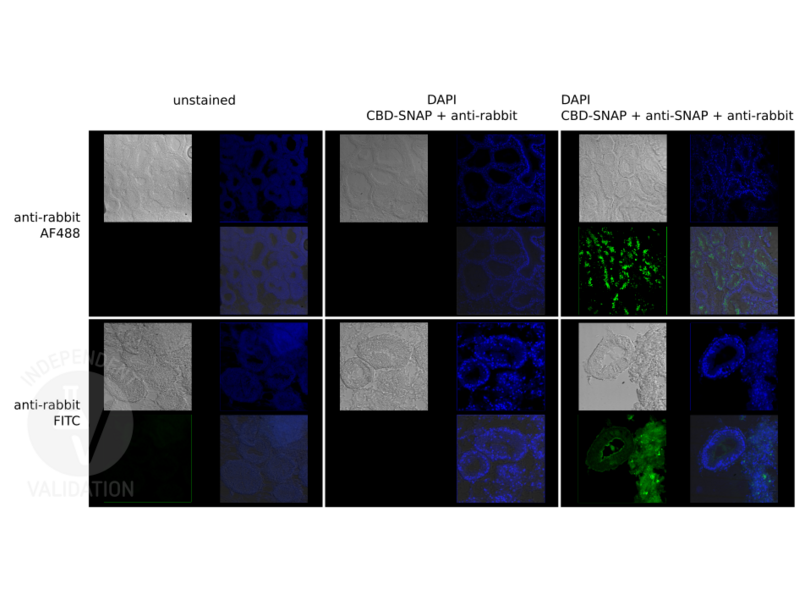
![Immunofluorescence images of oyster visceral mass tissue, with the specificity of Anti-Rabbit Alexa 488 (top row) and Anti-Rabbit FITC (bottom row) compared. Unstained samples (left column) were compared to test samples (right column) to visualize degree of auto-fluorescence. The absence of anti-SNAP served as the negative control (middle column), it has led to the absence of fluorescence at the same imaging and visualization setting for the green channels, implying the goat anti-rabbit IgG (Heavy & Light Chain) antibody FITC conjugate ABIN101988 is specific to Rabbit IgG antigen.]() Immunofluorescence images of oyster visceral mass tissue, with the specificity of Anti-Rabbit Alexa 488 (top row) and Anti-Rabbit FITC (bottom row) compared. Unstained samples (left column) were compared to test samples (right column) to visualize degree of auto-fluorescence. The absence of anti-SNAP served as the negative control (middle column), it has led to the absence of fluorescence at the same imaging and visualization setting for the green channels, implying the goat anti-rabbit IgG (Heavy & Light Chain) antibody FITC conjugate ABIN101988 is specific to Rabbit IgG antigen.
Full Methods
Immunofluorescence images of oyster visceral mass tissue, with the specificity of Anti-Rabbit Alexa 488 (top row) and Anti-Rabbit FITC (bottom row) compared. Unstained samples (left column) were compared to test samples (right column) to visualize degree of auto-fluorescence. The absence of anti-SNAP served as the negative control (middle column), it has led to the absence of fluorescence at the same imaging and visualization setting for the green channels, implying the goat anti-rabbit IgG (Heavy & Light Chain) antibody FITC conjugate ABIN101988 is specific to Rabbit IgG antigen.
Full Methods -
- Format
- Lyophilized
- Reconstitution
- Reconstitution Buffer: Restore with deionized water (or equivalent), Reconstitution Volume: 1.0 mL
- Concentration
- 2.0 mg/mL
- Buffer
-
Buffer: 0.02 M Potassium Phosphate, 0.15 M Sodium Chloride, pH 7.2
Stabilizer: 10 mg/mL Bovine Serum Albumin (BSA) - Immunoglobulin and Protease free
, Preservative:0.01 % (w/v) Sodium Azide - Preservative
- Sodium azide
- Precaution of Use
- This product contains Sodium azide: a POISONOUS AND HAZARDOUS SUBSTANCE which should be handled by trained staff only.
- Storage
- 4 °C,-20 °C
- Storage Comment
- Store vial at 4° C prior to restoration. For extended storage aliquot contents and freeze at -20° C or below. Avoid cycles of freezing and thawing. Centrifuge product if not completely clear after standing at room temperature. This product is stable for several weeks at 4° C as an undiluted liquid. Dilute only prior to immediate use.
- Expiry Date
- 12 months
-
-
: "Association between dietary selenium intake and the prevalence of osteoporosis and its role in the treatment of glucocorticoid-induced osteoporosis." in: Journal of orthopaedic surgery and research, Vol. 18, Issue 1, pp. 867, (2023) (PubMed).
: "Dexmedetomidine Confers Protection Against Neuronal Oxygen Glucose Deprivation-Reperfusion by Regulating SIRT3 Mediated Autophagy." in: Neurochemical research, Vol. 47, Issue 11, pp. 3490-3505, (2022) (PubMed).
: "MiR-200c/FUT4 axis prevents the proliferation of colon cancer cells by downregulating the Wnt/β-catenin pathway." in: BMC cancer, Vol. 21, Issue 1, pp. 2, (2021) (PubMed).
: "miR‑142‑3p targets AC9 to regulate sciatic nerve injury‑induced neuropathic pain by regulating the cAMP/AMPK signalling pathway." in: International journal of molecular medicine, Vol. 47, Issue 2, pp. 561-572, (2021) (PubMed).
: "Prognostic significance of BIRC7/Livin, Bcl-2, p53, Annexin V, PD-L1, DARC, MSH2 and PMS2 in colorectal cancer treated with FOLFOX chemotherapy with or without aspirin." in: PLoS ONE, Vol. 16, Issue 1, pp. e0245581, (2021) (PubMed).
: "LncRNA Neat1 Promotes Regeneration after Spinal Cord Injury by Targeting miR-29b." in: Journal of molecular neuroscience : MN, Vol. 71, Issue 6, pp. 1174-1184, (2021) (PubMed).
: "Effects of sevoflurane general anesthesia during early pregnancy on AIM2 expression in the hippocampus and parietal cortex of Sprague-Dawley offspring rats." in: Experimental and therapeutic medicine, Vol. 21, Issue 5, pp. 469, (2021) (PubMed).
: "IL-17 promotes proliferation, inflammation and inhibits apoptosis of HaCaT cells via interacting with the TRAF3 interacting protein 2." in: Experimental and therapeutic medicine, Vol. 21, Issue 1, pp. 49, (2021) (PubMed).
: "MicroRNA-29a promotes the proliferation of human nasal epithelial cells and inhibits their apoptosis and promotes the development of allergic rhinitis by down-regulating FOS expression." in: PloS one, Vol. 16, Issue 8, pp. e0255480, (2021) (PubMed).
: "Klotho Inhibits Proliferation in a RET Fusion Model of Papillary Thyroid Cancer by Regulating the Wnt/β-Catenin Pathway." in: Cancer management and research, Vol. 13, pp. 4791-4802, (2021) (PubMed).
: "Downregulation of miR‑146a inhibits osteoporosis in the jaws of ovariectomized rats by regulating the Wnt/β‑catenin signaling pathway." in: International journal of molecular medicine, Vol. 47, Issue 3, (2021) (PubMed).
: "miR-135a inhibits airway inflammatory response in asthmatic mice via regulating JAK/STAT signaling pathway." in: Brazilian journal of medical and biological research = Revista brasileira de pesquisas medicas e biologicas, Vol. 54, Issue 3, pp. e10023, (2021) (PubMed).
: "Sevoflurane Post-treatment Upregulated miR-203 Expression to Attenuate Cerebral Ischemia-Reperfusion-Induced Neuroinflammation by Targeting MyD88." in: Inflammation, (2020) (PubMed).
: "Resveratrol Plays Protective Roles on Kidney of Uremic Rats via Activating HSP70 Expression." in: BioMed research international, Vol. 2020, pp. 2126748, (2020) (PubMed).
: "Overexpression of miR-146a inhibits the apoptosis of hippocampal neurons of rats with cerebral hemorrhage by regulating autophagy." in: Human & experimental toxicology, pp. 960327120907131, (2020) (PubMed).
: "Dexmedetomidine Post-Conditioning Alleviates Cerebral Ischemia-Reperfusion Injury in Rats by Inhibiting High Mobility Group Protein B1 Group (HMGB1)/Toll-Like Receptor 4 (TLR4)/Nuclear Factor kappa B ..." in: Medical science monitor : international medical journal of experimental and clinical research, Vol. 26, pp. e918617, (2020) (PubMed).
: "Overexpression of microRNA-141 inhibits osteoporosis in the jawbones of ovariectomized rats by regulating the Wnt/β-catenin pathway." in: Archives of oral biology, Vol. 113, pp. 104713, (2020) (PubMed).
: "Dexmedetomidine Alleviates CCI-Induced Neuropathic Pain via Inhibiting HMGB1-Mediated Astrocyte Activation and the TLR4/NF-κB Signaling Pathway in Rats." in: Neurotoxicity research, (2020) (PubMed).
: "Formononetin inhibits inflammation and promotes gastric mucosal angiogenesis in gastric ulcer rats through regulating NF-κB signaling pathway." in: Journal of receptor and signal transduction research, pp. 1-7, (2020) (PubMed).
: "miR-124-3p Regulates FGF2-EGFR Pathway to Overcome Pemetrexed Resistance in Lung Adenocarcinoma Cells by Targeting MGAT5." in: Cancer management and research, Vol. 12, pp. 11597-11609, (2020) (PubMed).
-
: "Association between dietary selenium intake and the prevalence of osteoporosis and its role in the treatment of glucocorticoid-induced osteoporosis." in: Journal of orthopaedic surgery and research, Vol. 18, Issue 1, pp. 867, (2023) (PubMed).
-
- Target
- IgG
- Abstract
- IgG Products
- Target Type
- Antibody
- Background
- Secreted as part of the adaptive immune response by plasma B cells, immunoglobulin G constitutes 75 % of serum immunoglobulins. Immunoglobulin G binds to viruses, bacteria, as well as fungi and facilitates their destruction or neutralization via agglutination (and thereby immobilizing them), activation of the compliment cascade, and opsonization for phagocytosis. The whole IgG molecule possesses both the F(c) region, recognized by high-affinity Fc receptor proteins, as well as the F(ab) region possessing the epitope-recognition site. Both heavy and light chains of the antibody molecule are present. Secondary Antibodies are available in a variety of formats and conjugate types. When choosing a secondary antibody product, consideration must be given to species and immunoglobulin specificity, conjugate type, fragment and chain specificity, level of cross-reactivity, and host-species source and fragment composition. This Anti-Rabbit IgG (H&L) is conjugated to Fluorescein.
-


