TNF alpha ELISA Kit
-
- Target See all TNF alpha ELISA Kits
- TNF alpha (Tumor Necrosis Factor alpha (TNF alpha))
- Binding Specificity
- AA 77-233
-
Reactivity
- Human
- Detection Method
- Colorimetric
- Method Type
- Sandwich ELISA
- Detection Range
- 15.6 pg/mL - 1000 pg/mL
- Minimum Detection Limit
- 15.6 pg/mL
- Application
- ELISA
- Purpose
- Sandwich High Sensitivity ELISA kit for Quantitative Detection of Human TNF alpha
- Brand
- PicoKine™
- Sample Type
- Cell Culture Supernatant, Serum, Plasma (heparin), Plasma (EDTA), Plasma (citrate)
- Analytical Method
- Quantitative
- Specificity
- Expression system for standard: E.coli,V77-L233
- Cross-Reactivity (Details)
- There is no detectable cross-reactivity with other relevant proteins.
- Sensitivity
- <1pg/mL
- Material not included
- Microplate reader in standard size. Automated plate washer. Adjustable pipettes and pipette tips. Multichannel pipettes are recommended in the condition of large amount of samples in the detection. Clean tubes and Eppendorf tubes. Washing buffer (neutral PBS or TBS). Preparation of 0.01M TBS: Add 1.2g Tris, 8.5g Nacl
- Immunogen
-
Expression system for standard: E.coli
Immunogen sequence: V77-L233 - Top Product
- Discover our top product TNF alpha ELISA Kit
-
-
- Application Notes
- Before using Kit, spin tubes and bring down all components to bottom of tube. Duplicate well assay was recommended for both standard and sample testing.
- Comment
-
Sequence similarities: Belongs to the tumor necrosis factor family.
- Plate
- Pre-coated
- Protocol
- human TNF alpha ELISA Kit was based on standard sandwich enzyme-linked immune-sorbent assay technology. A monoclonal antibody from mouse specific for TNF alpha has been precoated onto 96-well plates. Standards (E.coli,V77-L233) and test samples are added to the wells, a biotinylated detection polyclonal antibody from goat specific for TNF alpha is added subsequently and then followed by washing with PBS or TBS buffer. Avidin-Biotin-Peroxidase Complex was added and unbound conjugates were washed away with PBS or TBS buffer. HRP substrate TMB was used to visualize HRP enzymatic reaction. TMB was catalyzed by HRP to produce a blue color product that changed into yellow after adding acidic stop solution. The density of yellow is proportional to the human TNF alpha amount of sample captured in plate.
- Assay Procedure
-
Aliquot 0.1 mL per well of the 1000pg/mL, 500pg/mL, 250pg/mL, 125pg/mL, 62.5pg/mL, 31.2pg/mL, 15.6pg/mL human TNF alpha standard solutions into the precoated 96-well plate. Add 0.1 mL of the sample diluent buffer into the control well (Zero well). Add 0.1 mL of each properly diluted sample of human cell culture supernates, serum or plasma(heparin, EDTA, citrate) to each empty well. See "Sample Dilution Guideline" above for details. It is recommended that each human TNF alpha standard solution and each sample be measured in duplicate.
- Assay Precision
-
- Sample 1: n=16, Mean(pg/ml): 93, Standard deviation: 5.1, CV(%): 5.5
- Sample 2: n=16, Mean(pg/ml): 327, Standard deviation: 15.4, CV(%): 4.7
- Sample 3: n=16, Mean(pg/ml): 608, Standard deviation: 31, CV(%): 5.1,
- Sample 1: n=24, Mean(pg/ml): 102, Standard deviation: 7.65, CV(%): 7.5
- Sample 2: n=24, Mean(pg/ml): 319, Standard deviation: 15.3, CV(%): 4.8
- Sample 3: n=24, Mean(pg/ml): 613, Standard deviation: 35, CV(%): 5.7
- Restrictions
- For Research Use only
-
- by
- Dept. of Radiation Oncology, Dana-Farber Cancer Institute, Harvard Medical School
- No.
- #102066
- Date
- 11/16/2017
- Antigen
- TNF
- Lot Number
- 23913851011
- Method validated
- ELISA
- Positive Control
- Four cell culture supernates of healthy human Peripheral Blood Mononuclear Cells (hPBMCs) stimulated by T-cell activator (anti CD3/CD28 Antibody). Healthy hPBMCs stimulated by T-cell activator are well known to release inflammatory cytokine such as TNFα and TNFγ. TNFα had been verified using a different ELISA before.
- Negative Control
- Complete medium using RPMI and human serum (10%)
- Notes
Passed. The human TNF ELISA kit ABIN411361 specifically recognizes TNFα in human PBMCs upon stimulation.
- Primary Antibody
- Secondary Antibody
- Full Protocol
- Harvest hPBMCs at different time points (before irradiation, 4h and 24h after irradiation) by centrifugation. Collect culture media and store them immediately at -20°C.
- Thaw samples on ice followed by centrifugation to remove any residual particulates.
- Reconstitute the human TNFα standard. Shake gently to avoid foaming.
- Prepare the biotinylated anti-Human TNFα antibody working solution 30min prior to the experiment according to the kit manual.
- Prepare the Avidin-Biotin-Peroxidase Complex (ABC) working solution 30min prior to the experiment according to the kit manual and keep it at 37°C until use.
- Keep the TMB color developing agent and TMB stop solution at 37°C for 30min until use.
- Prepare the Washing Buffer (0.01M PBS) according to the kit manual.
- Prepare the standards to make 1000pg/ml, 500pg/ml, 250pg/ml, 125pg/ml, 62.5pg/ml, 31.25pg/ml, 15.625pg/ml Human TNFα standard solutions: pipette 100µl of each of the standards, controls, and unknown samples into the assigned wells into the precoated 96-well plate. Add 100µl of the sample diluent buffer into the control well (Zero background well). Add 100µl of each sample to each empty well as indicated in figure panel A.
- Seal the plate with a new adhesive cover provided and incubate at 37°C for 90min.
- Remove the cover, discard the plate content, and blot the plate onto paper towels. The wells must not dry at any time.
- Add 100µl of biotinylated anti-Human TNFα antibody working solution into each well.
- Seal the plate with a new adhesive cover provided and incubate at 37°C for 60min.
- Remove the cover, discard the plate content, and wash the plate 3x for 1min with 0.01M PBS. Discard the washing buffer and blot the plate onto paper towels.
- Add 100µl of prepared ABC working solution into each well.
- Seal the plate with a new adhesive cover provided and incubate at 37°C for 30min.
- Wash the plate 5x for 1-2min with 0.01M TBS or 0.01M PBS. Discard the washing buffer and blot the plate onto paper towels.
- Add 90μl of prepared TMB color developing agent into each well.
- Seal the plate with a new adhesive cover and incubate at 37°C in dark for 20min.
- Add 100µl of prepared TMB stop solution into each well. The color changes into yellow immediately.
- Incubate for 10min after adding the stop solution.
- Read the OD absorbance at 450nm in a microplate reader within 30min after adding the stop solution.
- Experimental Notes
Samples were not diluted since a low target protein concentration was expected.
The standards and some samples were prepared in duplicates. The negative (n=1) and positive controls (n=4) were prepared in triplicates. The six different spike controls were measured without replicates (Figure panel A for details).
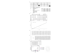
Increased TNFα release was detected in cell culture supernates stimulated by T cell activator without irradiation except for Sample #3. Low concentration of TNFα in all non-stimulated samples with or without irradiation were observed. We think that sample #3 was possibly a low responder for T cell stimulation (figure panel F).
Furthermore, in sample #1, TNFα level in cell culture supernate of irradiated cells (C-1) was slightly upregulated at 24 hours after irradiation compared with non-irradiated (C-3) (graph in figure panel F). Given this, although high-dose irradiation was probably enough to suppress TNFα release from the cells stimulated with T cell activator in some cells (samples #2 and #3), TNFα could be released by irradiation in some cells (sample #1).
Validation #102066 (ELISA)![Successfully validated 'Independent Validation' Badge]()
![Successfully validated 'Independent Validation' Badge]() Validation Images
Validation Images![TNFα measurements in human PBMCs using ABIN411361. Plate setup and nature of the samples (A). OD450 measurements and calculated concentrations of the utilized standards and controls (B). Standard curve generated using kit standards (C). OD450 measurements (D), calculated concentrations (E), and summary of sample measurements (F).]() TNFα measurements in human PBMCs using ABIN411361. Plate setup and nature of the samples (A). OD450 measurements and calculated concentrations of the utilized standards and controls (B). Standard curve generated using kit standards (C). OD450 measurements (D), calculated concentrations (E), and summary of sample measurements (F).
Full Methods
TNFα measurements in human PBMCs using ABIN411361. Plate setup and nature of the samples (A). OD450 measurements and calculated concentrations of the utilized standards and controls (B). Standard curve generated using kit standards (C). OD450 measurements (D), calculated concentrations (E), and summary of sample measurements (F).
Full Methods -
- Handling Advice
- Avoid multiple freeze-thaw cycles.
- Storage
- -20 °C,4 °C
- Storage Comment
- Store at 4°C for 6 months, at -20°C for 12 months. Avoid multiple freeze-thaw cycles
- Expiry Date
- 12 months
-
-
: "Mixture of Organophosphates Chronic Exposure and Pancreatic Dysregulations in Two Different Population Samples." in: Frontiers in public health, Vol. 8, pp. 534902, (2021) (PubMed).
: "4,4'-diaponeurosporene, a C30 carotenoid, effectively activates dendritic cells via CD36 and NF-κB signaling in a ROS independent manner." in: Oncotarget, Vol. 7, Issue 27, pp. 40978-40991, (2018) (PubMed).
: "LL202 protects against dextran sulfate sodium-induced experimental colitis in mice by inhibiting MAPK/AP-1 signaling." in: Oncotarget, Vol. 7, Issue 39, pp. 63981-63994, (2018) (PubMed).
: "An in vitro 3D bone metastasis model by using a human bone tissue culture and human sex-related cancer cells." in: Oncotarget, Vol. 7, Issue 47, pp. 76966-76983, (2018) (PubMed).
: "Proinflammatory effects of S100A8/A9 via TLR4 and RAGE signaling pathways in BV-2 microglial cells." in: International journal of molecular medicine, Vol. 40, Issue 1, pp. 31-38, (2018) (PubMed).
: "Heme Oxygenase-1, a Key Enzyme for the Cytoprotective Actions of Halophenols by Upregulating Nrf2 Expression via Activating Erk1/2 and PI3K/Akt in EA.hy926 Cells." in: Oxidative medicine and cellular longevity, Vol. 2017, pp. 7028478, (2018) (PubMed).
: "Wogonoside inhibits invasion and migration through suppressing TRAF2/4 expression in breast cancer." in: Journal of experimental & clinical cancer research : CR, Vol. 36, Issue 1, pp. 103, (2018) (PubMed).
: "Effect of different anesthetic methods on cellular immune functioning and the prognosis of patients with ovarian cancer undergoing oophorectomy." in: Bioscience reports, Vol. 37, Issue 5, (2018) (PubMed).
: "Essential Oil from Fructus Alpiniae Zerumbet Protects Human Umbilical Vein Endothelial Cells In Vitro from Injury Induced by High Glucose Levels by Suppressing Nuclear Transcription Factor-Kappa B ..." in: Medical science monitor : international medical journal of experimental and clinical research, Vol. 23, pp. 4760-4767, (2018) (PubMed).
: "Rottlerin as a therapeutic approach in psoriasis: Evidence from in vitro and in vivo studies." in: PLoS ONE, Vol. 12, Issue 12, pp. e0190051, (2018) (PubMed).
: "Salusin-β Is Involved in Diabetes Mellitus-Induced Endothelial Dysfunction via Degradation of Peroxisome Proliferator-Activated Receptor Gamma." in: Oxidative medicine and cellular longevity, Vol. 2017, pp. 6905217, (2018) (PubMed).
: "Inhibiting PSMα-induced neutrophil necroptosis protects mice with MRSA pneumonia by blocking the agr system." in: Cell death & disease, Vol. 9, Issue 3, pp. 362, (2018) (PubMed).
: "Safety of Low-calcium Dialysate and its Effects on Coronary Artery Calcification in Patients Undergoing Maintenance Hemodialysis." in: Scientific reports, Vol. 8, Issue 1, pp. 5941, (2018) (PubMed).
: "Expression of autophagy-related genes in cerebrospinal fluid of patients with tuberculous meningitis." in: Experimental and therapeutic medicine, Vol. 15, Issue 6, pp. 4671-4676, (2018) (PubMed).
: "Hypaphorine Attenuates Lipopolysaccharide-Induced Endothelial Inflammation via Regulation of TLR4 and PPAR-γ Dependent on PI3K/Akt/mTOR Signal Pathway." in: International journal of molecular sciences, Vol. 18, Issue 4, (2017) (PubMed).
: "Inflammatory cytokines and oxidative stress biomarkers in irritable bowel syndrome: Association with digestive symptoms and quality of life." in: Cytokine, Vol. 93, pp. 34-43, (2017) (PubMed).
: "Conditioned medium from relapsing-remitting multiple sclerosis patients reduces the expression and release of inflammatory cytokines induced by LPS-gingivalis in THP-1 and MO3.13 cell lines." in: Cytokine, Vol. 96, pp. 261-272, (2017) (PubMed).
: "Sulfated Cyclocarya paliurus polysaccharides markedly attenuates inflammation and oxidative damage in lipopolysaccharide-treated macrophage cells and mice." in: Scientific reports, Vol. 7, pp. 40402, (2017) (PubMed).
: "ox-LDL increases microRNA-29a transcription through upregulating YY1 and STAT1 in macrophages." in: Cell biology international, Vol. 41, Issue 9, pp. 1001-1011, (2017) (PubMed).
: "Associations of Trauma Severity with Mean Platelet Volume and Levels of Systemic Inflammatory Markers (IL1β, IL6, TNFα, and CRP)." in: Mediators of inflammation, Vol. 2016, pp. 9894716, (2017) (PubMed).
-
: "Mixture of Organophosphates Chronic Exposure and Pancreatic Dysregulations in Two Different Population Samples." in: Frontiers in public health, Vol. 8, pp. 534902, (2021) (PubMed).
-
- Target See all TNF alpha ELISA Kits
- TNF alpha (Tumor Necrosis Factor alpha (TNF alpha))
- Alternative Name
- TNF (TNF alpha Products)
- Background
-
Protein Function: Cytokine that binds to TNFRSF1A/TNFR1 and TNFRSF1B/TNFBR. It is mainly secreted by macrophages and can induce cell death of certain tumor cell lines. It is potent pyrogen causing fever by direct action or by stimulation of interleukin-1 secretion and is implicated in the induction of cachexia, Under certain conditions it can stimulate cell proliferation and induce cell differentiation. Impairs regulatory T-cells (Treg) function in individuals with rheumatoid arthritis via FOXP3 dephosphorylation. Upregulates the expression of protein phosphatase 1 (PP1), which dephosphorylates the key 'Ser-418' residue of FOXP3, thereby inactivating FOXP3 and rendering Treg cells functionally defective (PubMed:23396208). Key mediator of cell death in the anticancer action of BCG-stimulated neutrophils in combination with DIABLO/SMAC mimetic in the RT4v6 bladder cancer cell line (PubMed:22517918). .
Background: Tumor necrosis factor-alpha(TNF-alpha, or TNF) is secreted by macrophages in response to inflammation, infection and cancer. Human Tumor Necrosis Factor(TNF) and Lymphotoxin(TNF-beta) are cytotoxic proteins which have similar biological activities and share 30 % amino acid homology. TNF-alpha is produced by monocytes, which can stimulate endothelial cells to produce the multilineage growth factor granulocyte-macrophage colony-stimulating factor and extend the role of this immunoregulatory protein to the regulation of hematopoiesis in vitro. TNF is a soluble protein that causes damage to tumor cells but has no effect on normal cells. Human TNF has been purified to apparent homogeneity as a 17.3-kilodalton protein from HL-60 leukemia cells and has showed cytotoxic and cytostatic activities against various human tumor cell lines. The human TNF cDNA is 1585 base pairs in length and encodes a protein of 233 amino acids. The mature protein begins at residue 77, leaving a long leader sequence of 76 amino acids. TNF-alpha has been mapped to human chromosome 6.
Synonyms: Tumor necrosis factor,Cachectin,TNF-alpha,Tumor necrosis factor ligand superfamily member 2,TNF-a,Tumor necrosis factor, membrane form,N-terminal fragment,NTF,Intracellular domain 1,ICD1,Intracellular domain 2,ICD2,C-domain 1,C-domain 2,Tumor necrosis factor, soluble form,TNF,TNFA, TNFSF2,
Full Gene Name: Tumor necrosis factor
Cellular Localisation: Cell membrane, Single-pass type II membrane protein. - Gene ID
- 7124
- UniProt
- P01375
- Pathways
- NF-kappaB Signaling, Apoptosis, Caspase Cascade in Apoptosis, TLR Signaling, Cellular Response to Molecule of Bacterial Origin, Regulation of Leukocyte Mediated Immunity, Positive Regulation of Immune Effector Process, Production of Molecular Mediator of Immune Response, Positive Regulation of Endopeptidase Activity, Hepatitis C, Protein targeting to Nucleus, Inflammasome
-


 (158 references)
 (1 validation)


