MBL2 ELISA Kit
-
- Target See all MBL2 ELISA Kits
- MBL2 (Mannose-Binding Lectin (Protein C) 2, Soluble (MBL2))
-
Reactivity
- Human
- Detection Method
- Colorimetric
- Method Type
- Sandwich ELISA
- Detection Range
- 1.563-100 ng/mL
- Minimum Detection Limit
- 1.563 ng/mL
- Application
- ELISA
- Purpose
- For the quantitative determination of human mannma binding protein/mannan binding lectin (MBP/MBL) concentrations in serum, plasma and tissue homogenates.
- Sample Type
- Serum
- Analytical Method
- Quantitative
- Specificity
- This assay has high sensitivity and excellent specificity for detection of human MBP/MBL.
- Cross-Reactivity (Details)
- Limited by current skills and knowledge, it is impossible for us to complete the cross-reactivity detection between the target antigen and all analogues for other species. Therefore, cross reaction may still exist.
- Sensitivity
- 0.39 ng/mL
- Components
-
- Assay plate (12 × 8 coated Microwells)
- Standard (freeze dried)
- Biotin-antibody (100 × concentrate)
- HRP-avidin (100 × concentrate)
- Biotin-antibody Diluent
- HRP-avidin Diluent
- Sample Diluent
- Wash Buffer (25 × concentrate)
- TMB Substrate
- Stop Solution
- Adhesive Strip (for 96 wells)
- Instruction manual
-
-
- Application Notes
-
- The supplier is only responsible for the kit itself, but not for the samples consumed during the assay. The user should calculate the possible amount of the samples used in the whole test. Please reserve sufficient samples in advance.
- Samples to be used within 5 days may be stored at 2-8°C, otherwise samples must be stored at -20°C (≤ 1 month) or -80°C (≤ 2 months) to avoid loss of bioactivity and contamination.
- Grossly hemolyzed samples are not suitable for use in this assay.
- If the samples are not indicated in the manual, a preliminary experiment to determine the validity of the kit is necessary.
- Please predict the concentration before assaying. If values for these are not within the range of the standard curve, users must determine the optimal sample dilutions for their particular experiments.
- Tissue or cell extraction samples prepared by chemical lysis buffer may cause unexpected ELISA results due to the impacts of certain chemicals.
- Owing to the possibility of mismatching between antigens from another resource and antibodies used in this supplier's kits (e.g., antibody targets conformational epitope rather than linear epitope), some native or recombinant proteins from other manufacturers may not be recognized by this supplier's products.
- Influenced by factors including cell viability, cell number and cell sampling time, samples from cell culture supernatant may not be recognized by the kit.
- Fresh samples without long time storage are recommended for the test. Otherwise, protein degradation and denaturalization may occur in those samples and finally lead to wrong results.
- Comment
-
Detection wavelength: 450 nm
Information on standard material:
Depending on the antigen to be detected, standards can be either native or recombinant protein. The recombinant proteins are being expressed in CHO cells in most cases. Please inquire for more information. The formulation of auxiliary material in the standard is considered proprietary information, however it does not contain any poisonous substance. Proclin 300 (1:3000) is used as preservative.
Information on reagents:
In most cases the stop solution provided is 1 N H2SO4. The formulation of wash solution is proprietary information. None of the components contain (sodium) azide, thimerosal, 2-mercaptoethanol (2-ME) or any other poisonous materials. For the sandwich method kits, the sample diluent, antibody diluent, enzyme diluent and standard all contain BSA.
Information on antibodies:
The antibodies provided in different kits vary in regards to clonality and host. Some antibodies are affinity purified, some are Protein A - Sample Volume
- 100 μL
- Assay Time
- 1 - 4.5 h
- Plate
- Pre-coated
- Protocol
- This assay employs the quantitative sandwich enzyme immunoassay technique. Antibody specific for MBP/MBL has been pre-coated onto a microplate. Standards and samples are pipetted into the wells and any MBP/MBL present is bound by the immobilized antibody. After removing any unbound substances, a biotin-conjugated antibody specific for MBP/MBL is added to the wells. After washing, avidin conjugated Horseradish Peroxidase (HRP) is added to the wells. Following a wash to remove any unbound avidin-enzyme reagent, a substrate solution is added to the wells and color develops in proportion to the amount of MBP/MBL bound in the initial step. The color development is stopped and the intensity of the color is measured.
- Reagent Preparation
-
- Biotin-antibody (1×) - Centrifuge the vial before opening.
Biotin-antibody requires a 100-fold dilution. The suggested dilution is 10µL of Biotin-antibody + 990µL of Biotin-antibody Diluent. - HRP-avidin (1×) - Centrifuge the vial before opening.
HRP-avidin requires a 100-fold dilution. The suggested dilution is 10µL of HRP-avidin + 990µL of HRP-avidin Diluent. - Wash Buffer (1×) - If crystals have formed in the concentrate, warm up to room temperature and mix gently until the crystals have completely dissolved. Dilute 20mL of Wash Buffer Concentrate (25×) into deionized or distilled water to prepare 500mL of Wash Buffer (1×).
- Standard - Centrifuge the standard vial at 6000-10000rpm for 30s.
Reconstitute the Standard with 1ml of Sample Diluent. Do not substitute other diluents. This reconstitution produces a stock solution of 200pg/mL. Mix the standard to ensure complete reconstitution and allow the standard to sit for a minimum of 15 minutes with gentle agitation prior to making dilutions.
Pipette 250µL of Sample Diluent into each tube. Use the stock solution to produce a 2-fold dilution series. Mix each tube thoroughly before the next transfer. The undiluted Standard serves as the high standard (200pg/mL). Sample Diluent serves as the zero standard (0ng/mL).
- Kindly use graduated containers to prepare the reagent. Please don't prepare the reagent directly in the Diluent vials provided in the kit.
- Bring all reagents to room temperature (18-25°C) before use for 30 min.
- Prepare fresh standard for each assay. Use within 4 hours and discard after use.
- Making serial dilution in the wells directly is not permitted.
- Please carefully reconstitute Standards according to the instruction. Avoid foaming and mix gently until the crystals have completely dissolved. To minimize imprecision caused by pipetting, use small volumes and ensure that pipettors are calibrated. It is recommended to suck more than 10µL when pipetting.
- It is recommended to use distilled water to prepare reagents and samples. Using contaminated water or container for reagent preparation will influence detection result.
- Biotin-antibody (1×) - Centrifuge the vial before opening.
- Assay Precision
-
Intra-assay precision (precision within an assay): Three samples of known concentration were tested twenty times on one plate to assess precision.
Inter-assay precision (precision between assays): Three samples of known concentration were tested in twenty assays to assess precision.- Intra-assay: CV% less than 8%
- Inter-assay: CV% less than 10%
- Restrictions
- For Research Use only
-
- by
- CGIBD Advanced Analytics Core
- No.
- #029784
- Date
- 08/11/2014
- Antigen
- Lot Number
- Y25184072
- Method validated
- ELISA
- Positive Control
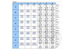
- Human serum - expression is ~50 ng/mL
- Negative Control
- Goat serum (non-reactive species)
- Notes
- Target protein was detected in the positive control sample and not in the negative control sample as expected.
- Primary Antibody
- Secondary Antibody
- Full Protocol
- 1. All reagents were brought up to room temperature for 30 minutes prior to use. The 1x Wash Buffer was prepared by adding 20 mL of 25x Wash Buffer Concentrate to 480 mL of distilled/deionized water and mixing thoroughly.
- 2. The vial of Standard was reconstituted with 1 mL of Sample Diluent, mixed, and allowed to sit for 15 minutes with gentle agitation.
- 3. The standard curve was prepared by creating a 2-fold dilution series of seven standards (including the original undiluted vial) using Sample Diluent. Sample Diluent alone served as the 0 pg/mL standard.
- 4. The assay plate was removed from the foil pouch and 100 μL of each standard and sample were added to the appropriate wells, in triplicate. The plate was covered with the adhesive strip provided and incubated for 2 hours at 37 °C.
- 5. Approximately 10 minutes before the incubation ended, a 1x Biotin-antibody solution was prepared by diluting 60 μL of 100x Biotin-antibody into 5940 μL of Biotin-antibody Diluent.
- 6. The liquid from each well was removed.
- 7. 100 μL of 1x Biotin-antibody solution was added to each well, and the plate was covered with a new adhesive strip, and incubated for 1 hour at 37 °C.
- 8. Approximately 10 minutes before the incubation ended, a 1x HRP-avidin solution was prepared by diluting 60 μL of 100x HRP-avidin into 5940 μL of HRP-avidin Diluent.
- 9. Each well was aspirated and washed, repeating the process two times for a total of three washes. Each well was washed by filling each well with 1x Wash Buffer and letting it stand for 2 minutes. After the last wash, remaining Wash Buffer was removed and the plate was inverted and blotted against clean, absorbent paper towels.
- 10. 100 μL of 1x HRP-avidin solution was added to each well, the plate was covered with a new adhesive strip, and incubated for 1 hour at 37 °C.
- 11. The aspiration/wash procedure from Step 9 was repeated for an additional 5 washes.
- 12. 90 μL of TMB Substrate was added to each well. The plate was protected from light and incubated for 15-30 minutes at 37 °C, with periodic checking to prevent overdevelopment.
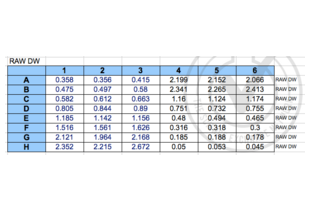
- 13. 50 μL of Stop Solution was added to each well and mixed thoroughly. The optical density (OD) of each well was measured within 5 minutes using a microplate reader set to 450 nm.
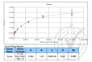
- 14. A standard curve was generated by plotting the OD value for each standard on the y-axis against the concentration on the x-axis. A line of best fit through the points on the graph was used to generate an equation to calculate MBP concentrations of the samples based on their average OD values.
- Experimental Notes
- The 100x HRP-avidin provided contained less than 60 μL. Other than that, there were no experimental challenges noted.
Validation #029784 (ELISA)![Successfully validated 'Independent Validation' Badge]()
![Successfully validated 'Independent Validation' Badge]() Validation Images
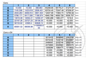
Validation Images![Figure 4: MBP concentrations calculated from standard curve formula. Upper panel = uncorrected for dilution; lower panel = corrected for dilution. On average, 3490315 pg/mL of MBP was detected in the positive control (human serum) and 0 pg/mL of MBP was detected in the negative control (goat serum).]() Figure 4: MBP concentrations calculated from standard curve formula. Upper panel = uncorrected for dilution; lower panel = corrected for dilution. On average, 3490315 pg/mL of MBP was detected in the positive control (human serum) and 0 pg/mL of MBP was detected in the negative control (goat serum).
Full Methods
Figure 4: MBP concentrations calculated from standard curve formula. Upper panel = uncorrected for dilution; lower panel = corrected for dilution. On average, 3490315 pg/mL of MBP was detected in the positive control (human serum) and 0 pg/mL of MBP was detected in the negative control (goat serum).
Full Methods -
- Precaution of Use
- The Stop Solution provided with this kit is an acid solution. Wear eye, hand, face and clothing protection when using this material.
- Handling Advice
-
- The kit should not be used beyond the expiration date on the kit label.
- Do not mix or substitute reagents with those from other lots or sources.
- If samples generate values higher than the highest standard, dilute the samples with Sample Diluent and repeat the assay.
- Any variation in Sample Diluent, operator, pipetting technique, washing technique, incubation time/temperature and kit age can cause variation in binding.
- This assay is designed to eliminate interference by soluble receptors, binding proteins and other factors present in biological samples. Until all factors have been tested in the Immunoassay, the possibility of interference cannot be excluded.
- Storage
- 4 °C/-20 °C
- Storage Comment
- For unopened kit: All the reagents should be kept according to the labels on vials.
- Expiry Date
- 6 months
-
-
: "Prognostic value of mannose-binding lectin: 90-day outcome in patients with acute ischemic stroke." in: Molecular neurobiology, Vol. 51, Issue 1, pp. 230-9, (2015) (PubMed).
: "Low levels of mannose-binding lectin at admission increase the risk of adverse neurological outcome in preterm infants: a 1-year follow-up study." in: The journal of maternal-fetal & neonatal medicine, pp. 1-5, (2015) (PubMed).
-
: "Prognostic value of mannose-binding lectin: 90-day outcome in patients with acute ischemic stroke." in: Molecular neurobiology, Vol. 51, Issue 1, pp. 230-9, (2015) (PubMed).
-
- Target See all MBL2 ELISA Kits
- MBL2 (Mannose-Binding Lectin (Protein C) 2, Soluble (MBL2))
- Alternative Name
- mannose-binding lectin (protein C) 2, soluble (opsonic defect) (MBL2 Products)
- Background
- Synonyms: COLEC1, HSMBPC, MBL, MBP, MBP1, MGC116832, MGC116833, mannan-binding lectin|mannose-binding lectin 2, soluble (opsonic defect)|mannose-binding protein C|soluble mannose-binding lectin
- HGNC
- 6922
- UniProt
- P11226
- Pathways
- Complement System, Positive Regulation of Immune Effector Process
-

 (2 references)
 (1 validation)