alpha Fetoprotein ELISA Kit
-
- Target See all alpha Fetoprotein (AFP) ELISA Kits
- alpha Fetoprotein (AFP) (alpha-Fetoprotein (AFP))
-
Reactivity
- Human
- Detection Method
- Colorimetric
- Method Type
- Sandwich ELISA
- Detection Range
- 1.56 ng/mL - 100 ng/mL
- Minimum Detection Limit
- 1.56 ng/mL
- Application
- ELISA
- Purpose
- The kit is a sandwich enzyme immunoassay technique for the in vitro quantitative measurement in various sample types.
- Sample Type
- Plasma, Serum
- Analytical Method
- Quantitative
- Specificity
- This kit recognizes natural and recombinantHumanαFP. No significant cross-reactivity or interference between HumanαFP and analogues was observed. Note: Limited by existing techniques, cross reaction may still exist, as it is impossible for us to complete the cross-reactivity detection between HumanαFP and all the analogues.
- Cross-Reactivity (Details)
- No significant cross-reactivity or interference between human Alpha-Fetoprotein and analogues was observed. Note: Limited by current skills and knowledge, it is impossible for us to complete the cross-reactivity detection between Human bFGF and all the analogues, therefore, cross reaction may still exist.
- Sensitivity
- 0.94 ng/mL
- Components
-
- Pre-coated, ready to use 96-well strip plate, flat buttom
- Plate sealer for 96 wells
- Reference Standard
- Reference Standard & Sample Diluent
- Biotinylated Detection Antibody (100 x concentrate)
- HRP Conjugate (100 x concentrate)
- Biotinylated Detection Antibody Diluent
- HRP Conjugate Diluent
- Substrate Reagent
- Stop Solution
- Wash Buffer (25 x concentrate)
- Instruction manual
- Top Product
- Discover our top product AFP ELISA Kit
-
-
- Application Notes
-
ELISA Plate: The just opened ELISA Plate may appear water-like substance, which is normal and will not have any impact on the experiment results.
Add Sample: The interval of sample adding between the first well and the last well should not be too long, otherwise will cause different pre-incubation time, which will significantly affect the experiment’s accuracy and repeatability. For each step in the procedure, total dispensing time for addition of reagents or samples to the assay plate should not exceed 10 minutes. Parallel measure ment is recommended.
Incubation: To prevent evaporation and ensure accurate results, proper adhesion of plate sealers during incubation steps is necessary. Do not allow wells to sit uncovered for extended periods between incubation steps. Do not let the strips dry at any time during the assay. Strict compliance with the given incubation time and temperature.
Washing: The wash procedure is critical. Insufficient washing will result in poor precision and falsely elevated absorbance readings. Residual liquid in the reaction wells should be pat dry against absorbent paper in the washing process. But don’t put absorbent paper into reaction wells directly. Note that clear the residual liquid and fingerprint in the bottom before measurement, so as not to affect the micro-titer plate reader.
Reagent Preparation: As the volume of Detection Ab and HRP Conjugate is very small, liquid may adhere to the tube wall or tube cap when being transported. You better hand-throw it or centrifugal it for 1 minute at 1000rpm. Please pipette the solution for 4-5 times before pippeting. Please carefully reconstitute Standards, working solutions of Detection Ab and HRP Conjugate according to the instructions. To minimize imprecision caused by pipetting, ensure that pipettors are calibrated. It is recommended to suck more than 10μL for once pipetting. Do not reuse standard solution, working solution of Detection Ab and HRP Conjugate, which have been diluted. If you need to use standard repeatedly, you can divide the standard into small pack according to the amount of each assay, keep them at -20°C to -80°C and avoid repeated freezing and thawing.
Reaction Time Control: Please control reaction time strictly following this product description!
Substrate: Substrate Solution is easily contaminated. Please protect it from light.Stop Solution: As it is an acid solution, please pay attention to the protection of your eyes, hands, face and clothes when using this solution.
Mixing: You’d better use micro-oscillator at the lowest frequency, as sufficient and gentle mixing is particularly important to reaction result. If there is no micro-oscillator available, you can knock the ELISA plate frame gently with your finger before reaction.
Security: Please wear lab coats and latex gloves for protection. Especially detecting samples of blood or other body fluid, please perform following the national security columns of biological laboratories.
Do not use component from different batches of kit(washing buffer and stop solution can be an exception)
To avoid cross-contamination, change pipette tips between adding of each standard level, between sample adding, and between reagent adding. Also, use separate reservoirs for each reagent. Otherwise, the results will be inaccurate! - Comment
-
Information on standard material:
The formulation of the standard is 0.01 M PBS. The standard contains additives (1 % BSA).
Information on reagents:
Reagents include 1 M SO2. Azide, thimerosal, 2-mercaptoethanol (2-ME) or any other poisonous materials are not used.
Information on antibodies:
The provided antibodies and their host vary in different kits. All antibodies are affinity purified
The sensitivity of this assay, or Lower Limit of Detection (LLD) was defined as the lowest protein concentration that could be differentiated from zero. It was determined by adding two standard deviations to the mean optical density value of twenty zero standard replicates and calculating the corresponding concentration. - Sample Volume
- 100 µL
- Plate
- Pre-coated
- Protocol
-
- Add 100 µL standard or sample to each well. Incubate for 90 min at 37 °C.
- Remove the liquid. Add 100 µL Biotinylated Detection Antibody. Incubate for 1 hour at 37 °C.
- Aspirate and wash 3 times.
- Add 100 µL HRP Conjugate. Incubate for 30 min at 37 °C.
- Aspirate and wash 5 times.
- Add 90 µL Substrate Reagent. Incubate for 15 min at 37 °C.
- Add 50 µL Stop Solution. Read at 450 nm immediately.
- Calculation of results.
- Reagent Preparation
-
- Bring all reagents to room temperature (18~25 °C) before use. Follow the Microplate reader manual for set-up and preheat it for 15 min before OD measurement.
- Wash Buffer: Dilute 30 mL of Concentrated Wash Buffer with 720 mL of deionized or distilled water to prepare 750 mL of Wash Buffer.Note: if crystals have formed in the concentrate, warm it in a 40 °C water bath and mix it gently until the crystals have completely dissolved
- Standard working solution: Centrifuge the standard at 10,000xg for 1 min. Add 1.0 mL of Reference Standard &Sample Diluent, let it stand for 10 min and invert it gently several times. After it dissolves fully, mix it thoroughly with a pipette. This reconstitution produces a working solution of 100 ng/mL. Then make serial dilutions as needed. The recommended dilution gradient is as follows: 100, 50, 25, 12.5, 6.25, 3.13, 1.56, 0 ng/mL. Dilution method: Take 7 EP tubes, add 500 μLof Reference Standard & Sample Diluent to each tube. Pipette 500 μLof the 100 ng/mL working solution to the first tube and mix up to produce a 50 ng/mL working solution. Pipette 500 μLof the solution from the former tube into the latter one according to these steps. The illustration below is for reference. Note: the last tube is regarded as a blank. Don't pipette solution into it from the former tube.
- Biotinylated Detection Antibody working solution: Calculate the required amount before the experiment (100 μL/well). In preparation, slightly more than calculated should be prepared. Centrifuge the stock tube before use, dilute the 100x Concentrated Biotinylated Detection Antibody to 1xworking solution with Biotinylated Detection Antibody Diluent.
- Concentrated HRP Conjugate working solution: Calculate the required amount before the experiment (100 μL/well). In preparation, slightly more than calculated should be prepared. Dilute the 100x Concentrated HRP Conjugate to 1x working solution with Concentrated HRP Conjugate Diluent.
- Restrictions
- For Research Use only
-
- by
- Proteopath Clinical Lab
- No.
- #029781
- Date
- 07/29/2014
- Antigen
- Lot Number
- AK0014APR22014
- Method validated
- ELISA
- Positive Control
- Human plasma - expression is 3 ng/mL
- Negative Control
- Rabbit plasma (non-reactive species), mouse plasma (non-reactive species)
- Notes
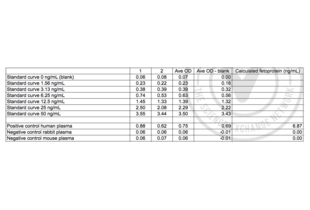
- Target protein was detected in the positive control sample (human) and not in the negative control samples (rabbit and mouse), as expected.
- Primary Antibody
- Secondary Antibody
- Full Protocol
- 1. All reagents were brought up to room temperature prior to use. The 1x Wash Buffer was prepared by adding 30 mL of Concentrated Wash Buffer into 750 mL of distilled/deionized water and mixing thoroughly.
- 2. The vial of Standard was reconstituted with 1 mL of Sample Diluent, mixed, and allowed to sit for 10 min with gentle agitation.
- 3. The standard curve was prepared by creating a 2-fold dilution series of seven standards (including the original undiluted vial) using Sample Diluent. Sample Diluent alone served as the 0 ng/mL standard.
- 4. The Biotinylated Detection Ab was prepared by diluting the concentrated stock 1:100 in Biotinylated Detection Ab Diluent.
- 5. The HRP Conjugate was prepared by diluting the concentrated stock 1:100 in HRP Conjugate Diluent.
- 6. 100 μL of each standard and sample were added per well to the ELISA plate in duplicate and mixed gently. The plate was covered with the sealer provided and incubated for 90 min at 37 °C.
- 7. The liquid from each well was removed.
- 8. 100 μL of Biotinylated Detection Ab working solution was added to each well, and the plate was covered with sealer, gently mixed, and incubated for 1 hour at 37 °C.
- 9. Each well was aspirated and washed, repeating the process two times for a total of three washes. Each well was washed by filling each well with 350 μL of 1x Wash Buffer and letting it stand for 2 minutes. After the last wash, remaining Wash Buffer was removed and the plate was inverted and blotted against clean, absorbent paper towels.
- 10. 100 μL of HRP conjugate working solution was added to each well, the plate was covered with sealer, and incubated for 30 min at 37 °C.
- 11. The aspiration/wash procedure from Step 9 was repeated for an additional 5 washes.
- 12. 90 μL of Substrate Solution was added to each well and the plate was covered with a new plate sealer. The plate was protected from light and incubated for 15 min at 37 °C, with periodic checking to prevent overdevelopment.
- 13. 50 μL of Stop Solution was added to each well and mixed thoroughly. The optical density (OD) of each well was measured immediately using a microplate reader set to 450 nm.
- 14. The duplicate readings for each standard and samples were averaged, and the average zero (blank) standard optical density was subtracted from each value.
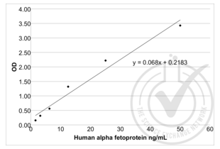
- 15. A standard curve was generated by plotting the blanked OD value for each standard on the y-axis against the concentration on the x-axis using Excel. A line of best fit through the points on the graph was used to generate the equation x = (y-0.2183) / 0.068.
- 16. The equation was used to calculate AFP concentrations of the samples based on their average blanked OD values.
- Experimental Notes
- Nothing to note
Validation #029781 (ELISA)![Successfully validated 'Independent Validation' Badge]()
![Successfully validated 'Independent Validation' Badge]() Validation Images
Validation Images![Figure 2: Table of absorbance readings (OD 450 nm) for standard curve, positive (human plasma) and negative (rabbit, mouse plasma) control samples. Value for Average Reading is derived from the average of two readings (OD 450nm). The average of the blank was subtracted from all readings and the average blanked OD readings were used to plot a standard curve. An equation x = (y - 0.2183) / 0.068 was generated from the standard curve and used to calculate alpha-Fetoprotein (AFP) concentrations shown in the Table.]() Figure 2: Table of absorbance readings (OD 450 nm) for standard curve, positive (human plasma) and negative (rabbit, mouse plasma) control samples. Value for Average Reading is derived from the average of two readings (OD 450nm). The average of the blank was subtracted from all readings and the average blanked OD readings were used to plot a standard curve. An equation x = (y - 0.2183) / 0.068 was generated from the standard curve and used to calculate alpha-Fetoprotein (AFP) concentrations shown in the Table.
Full Methods
Figure 2: Table of absorbance readings (OD 450 nm) for standard curve, positive (human plasma) and negative (rabbit, mouse plasma) control samples. Value for Average Reading is derived from the average of two readings (OD 450nm). The average of the blank was subtracted from all readings and the average blanked OD readings were used to plot a standard curve. An equation x = (y - 0.2183) / 0.068 was generated from the standard curve and used to calculate alpha-Fetoprotein (AFP) concentrations shown in the Table.
Full Methods -
- Handling Advice
- All the reagents in the kit should be stored according to the labels on vials. Unused wells should be returned to the foil pouch with the desiccant pack and resealed along entire edge of zip-seal. Substrate Reagent shouldn't be kept at -20 °C (Check!). Exposure of reagents to strong light should be avoided in the process of incubation and storage. All the taps of reagents should be tightened to prevent evaporation and microbial contamination. If not to store reagents according to above suggestions, erroneous results may occur.
- Storage
- 4 °C/-20 °C
- Storage Comment
- The unopened kit can be stored at 4℃ for 1 month. If the kit is not used within 1 month, store the items separately according to the conditions since the kit is received.
-
- Target See all alpha Fetoprotein (AFP) ELISA Kits
- alpha Fetoprotein (AFP) (alpha-Fetoprotein (AFP))
- Alternative Name
- Alpha-Fetoprotein (AFP Products)
- Background
- Synonyms: AFP, FETA, HPAFP, Alpha-fetoglobulin, oralpha fetal protein
- Pathways
- C21-Steroid Hormone Metabolic Process
-


 (1 validation)


