Fluorescent Western Blotting
Fluorescent Western blotting can offer many advantages and improve the highly robust Western blotting technique. Fluorescent Western blotting is a method that is becoming increasingly popular because it allows powerful quantitative detection of protein expression.
The basic principle behind the method is that the strength of fluorescence of a fluorophore bound to a detection antibody is proportional to the strength of protein expression. Since fluorescence does not occur enzymatically, as is the case with classical Western blotting, quantitation is much more reproducible.
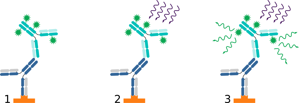
Figure 1. Fluorescent Westernblotting: 1. The fluorophore-conjugated secondary recognizes the primary antibody bound to the protein of interest. 2. A light source is used to excite the fluorescent conjugate to fluorescence. 3. The excited dye emits light of a specific wavelength, which can be visualized by digital imaging. Thus, the protein of interest can be detected quantitatively.
Fluorescence vs Chemiluminescence Western Blotting
| Chemiluminescent detection | Fluorescent detection | |
|---|---|---|
| Signal source | Signal from enzymatic reaction | Signal from fluorophore |
| Detection | Photographic film, imaging instrument | Imaging instrument with light source and filters |
| Sensitivity | Good, wide variety of substrates available | Good, wide variety of fluorophores available |
| Quantitation | Single channel, Challenging normalization | Multiple channel, easier normalization |
| Signal duration | Minutes to hours | Weeks to months |
Blocking Buffer for Fluorescent Western blotting
- Western Blot Blocking Solution is specifically designed for western blotting using fluorochrome conjugated antibodies.
- Aseptically filtered through a Millipore 0.22 micron filter into clean, pre-sterilized containers.
- Suitable for following western blot imaging systems: Bio-Rad Laboratories, GE Healthcare, Alpha Innotech, FujiFilm Life Science, Licor Biosciences, UVP and Syngene.
- In stock, fast delivery
- Western Blot Blocking Buffer is ideal for infrared Western Blotting.
- This product is a 2X concentrated stock solution.
- Prepare a 1X working solution by diluting 1 part 2X concentrate with 1 parts TBS or equivalent.
- Aseptically filtered through a Millipore 0.22 micron filter.
- In stock, fast delivery
Multiplex Duo Fluorescent Western Blotting Kits
Multiplex Duo fluorescent Western blotting kits are suited for simultaneous detection and quantification of specific protein populations in a biological sample. Using a combination of two antibodies selected for minimal cross reactivity, fluorescent detection method enables simultaneous quantitative analysis of multiple proteins within the same sample on the same blot.
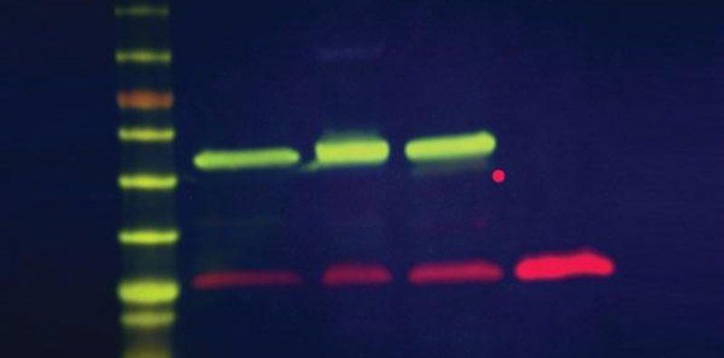
Figure 2. Multiplex Duo Fluorescent Western Blotting Kits: Simultaneous detection of α-tubulin and GFP on a single blot using labeled secondary antibody conjugates. Probing of cell lysates and GFP with mouse anti-α-tubulin and chicken anti-GFP antibodies followed by 649 goat anti-mouse IgG (pseudocolored green) and 800 goat anti-chicken IgG (red) conjugates, and then imaged using Syngene G:BOX Imaging System resulted in comparable patterns of detection.
| Name / Product link | Fluorescence channels | Reactivity |
|---|---|---|
| DyLight™ Multiplex 488/800 Duo Western Blot Kit | 488 / 800 nm | Mouse, Rabbit |
| DyLight™ Multiplex 549/800 Duo Western Blot Kit | 549 / 800 nm | Mouse, Rabbit |
| DyLight™ Multiplex 649/488 Duo Western Blot Kit | 649 / 488 nm | Mouse, Rabbit |
| DyLight™ Multiplex 649/549 Duo Western Blot Kit | 649 / 549 nm | Mouse, Rabbit |
| DyLight™ Multiplex 649/800 Duo Western Blot Kit | 649 / 800 nm | Mouse, Rabbit |
Multiplex Trio Fluorescent Western Blot Kits
The multiplex Trio fluorescent Western blot kit is suited for simultaneous detection and quantification of specific protein populations in a biological sample. Using a combination of three antibodies selected for minimal cross reactivity, fluorescent detection method enables simultaneous quantitative analysis of multiple proteins within the same sample on the same blot.
| Name / Product link | Fluorescence channels | Reactivity |
|---|---|---|
| DyLight™ Multiplex 649/488/800 Trio Western Blot Kit | 649 / 488 / 800 nm | Chicken, Mouse, Rabbit |
| DyLight™ Multiplex 649/549/800 Trio Western Blot Kit | 649 / 549 / 800 nm | Chicken, Mouse, Rabbit |
DyLight™ conjugated Secondary Antibodies
TrueBlot® HRP-conjugated Secondary Antibodies
antibodies-online.com: Making the Complex Convenient
- Target Coverage: Antibodies, proteins, kits and lysates covering a wide range of relevant targets
- Product Search: Extensive filtering options and comprehensive product details leading to the right choice
- Order Fulfillment: Trusted Supplier to 10000+ Institutions with Customer Service Hubs in US and Europe

Master of Science in engineering. 12+ years of experience in marketing and e-commerce in the life science sector.
Go to author page


