beta Actin antibody
 (2 references)
 (1 validation)
-
- Target See all beta Actin (ACTB) Antibodies
- beta Actin (ACTB) (Actin, beta (ACTB))
-
Reactivity
- Human, Mouse, Chicken, Rabbit, Pig, Cow, Hamster, Goat, Fish
-
Host
- Mouse
-
Clonality
- Monoclonal
-
Conjugate
- This beta Actin antibody is un-conjugated
-
Application
- Western Blotting (WB), ELISA
- Specificity
- THETM beta Actin Antibody, mAb, Mouse reacts with mouse, rabbit, chicken, human, hamster, cow, goat, fish, and pig. It has not yet been tested in other species.
- Cross-Reactivity (Details)
- Cross reactivity in other species has not yet been tested.
- Purification
- Protein G affinity column
- Immunogen
- A synthetic peptide DDDIAALVVDNGSG coupled - KLH
- Clone
- 2D1D10
- Isotype
- IgG
- Top Product
- Discover our top product ACTB Primary Antibody
-
anti-Actin, beta (ACTB) (AA 1-50) antibody
ACTB Reactivity: Human, Mouse, Rat, Rabbit, Pig, Cow WB, ELISA, IHC (p), FACS, IF (p), IF (cc) Host: Rabbit Polyclonal unconjugated
anti-Actin, beta (ACTB) (AA 2-16) antibodyACTB Reactivity: Mouse, Rat, Chicken, Zebrafish (Danio rerio) WB, IHC, ICC Host: Rabbit Polyclonal unconjugated
anti-Actin, beta (ACTB) antibodyACTB Reactivity: Human, Mouse, Rat WB, IF, IHC (p), FACS, DNA Mic Host: Mouse Monoclonal 137CT26-1-1 unconjugated
anti-Actin, beta (ACTB) (AA 350-375) antibodyACTB Reactivity: Human, Mouse, Leopard Frog WB, ELISA, IF, FM Host: Rabbit Polyclonal unconjugated
anti-Actin, beta (ACTB) (AA 1-25) antibodyACTB Reactivity: Human, Mouse, Rat, Pig, Cow WB, IHC (p) Host: Mouse Monoclonal 1A2 unconjugated
anti-Actin, beta (ACTB) (AA 1-375) antibodyACTB Reactivity: Human WB, ELISA, IHC (p) Host: Mouse Monoclonal 3G4-F9 unconjugated
anti-Actin, beta (ACTB) (N-Term) antibodyKO Validated ACTB Reactivity: Human WB, IF, IHC (p), ICC Host: Rabbit Polyclonal unconjugated
anti-Actin, beta (ACTB) (AA 1-50) antibodyACTB Reactivity: Human WB, ELISA, IHC, IF, IP, FACS Host: Mouse Monoclonal 1D12E12B5 unconjugated
anti-Actin, beta (ACTB) (N-Term) antibodyACTB Reactivity: Human, Mouse, Rat, Chicken, Rabbit, Dog, Monkey, Pig, Cow, Sheep, Hamster, Horse, Guinea Pig, Goat, Cat, Avian, Donkey WB, IF Host: Goat Polyclonal unconjugated
anti-Actin, beta (ACTB) (Middle Region) antibodyACTB Reactivity: Human, Mouse, Rat, Pig, Cow, Sheep, Zebrafish (Danio rerio), Horse, Goat WB, IHC Host: Rabbit Polyclonal unconjugated
-
- Application Notes
-
Working concentrations for specific applications should be determined by the investigator. The appropriate concentrations may be affected by secondary antibody affinity, antigen concentration, the sensitivity of the method of detection, temperature, the length of the incubations, and other factors. The suitability of this antibody for applications other than those listed below has not been determined. The following concentration ranges are recommended starting points for this product.
ELISA: 0.1-1.0 µg/mL
Western blot: 1.0 µg/mL
Other applications: user-optimized - Restrictions
- For Research Use only
-
- by
- Dr. Randy Brutkiewicz Laboratory, Department of Microbiology and Immunology, Indiana University School of Medicine
- No.
- #102832
- Date
- 02/20/2018
- Antigen
- ACTB
- Lot Number
- Method validated
- Western Blotting
- Positive Control
- lysates from HEK293 cells untransfected or transfected with human MR1 cDNA
- Negative Control
- Notes
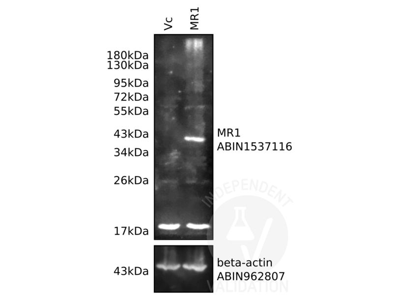
Passed. ABIN962807 recognizes human beta actin in HEK293 cell lysates.
- Primary Antibody
- ABIN962807
- Secondary Antibody
- goat anti-rabbit Dye-IR800 conjugated antibody (Advansta, R-05060-250, lot 17083179)
- Full Protocol
- Grow HEK293 cells in DMEM medium (Lonza, 12-614F, lot 0000618582) supplemented with serum (Hyclone, SH30071.03, lot AAG205460) and antibiotics (Hyclone, SV30010, lot J150013), at 37°C and 5% CO2 dish to 70-90% confluency.
- Transfect cells with pCDNA 3.1 neo (-) (Invitrogen) containing human MR1 cDNA (Genecopoeia) using Polyethylenimine (Polysciences, 23966) following the manufacturer´s instructions.
- Lyse cells in cold lysis buffer (10mM Tris pH7.4, 150mM NaCl, 0.5mM EDTA, 2% CHAPS).
- Determine total protein content of the lysates using Commassie Protein Assay Reagent (Thermo Scientific, 1856209, lot NL179252).
- Denature 200µg total protein for 5min at 95°C in 20µl Laemmli SDS sample buffer and subsequently separate them on a SDS-PAGE gel using Acrylamide/Bis Premixed (Bio-Rad, 61-0125, lot 260000477) for 2-3h at 100V.
- Transfer proteins onto PVDF membrane (Millipore, IPVH00010, lot K5AA6843U) with a Western blotting system for ON at 4°C at 150mA.
- Block the membrane with blocking buffer (2% BSA/PBS/0.05%Tween-20) for 1h at RT.
- Incubate membrane with:
- loading control rabbit anti beta-actin (antibodies-online, ABIN962807) diluted 1:500 in blocking buffer ON at 4°C.
- primary rabbit anti-MR1 antibody (antibodies-online, ABIN1537116, lot SA111213CH) diluted 1:1000 in blocking buffer ON at 4°C.
- Wash membrane 3x for 10min with PBS/0.05%Tween-20.
- Incubate membrane with secondary goat anti-rabbit Dye-IR800 conjugated antibody (Advansta, R-05060-250, lot 17083179) diluted 1:10000 in PBS/0.05% Tween-20 for 1h at RT.
- Wash membrane 3x for 10min with PBS/0.05% Tween-20.
- Reveal protein bands using an Odyssey imaging system (LI-COR Biosciences).
- Experimental Notes
The beta Actin antibody ABIN962807 reveals a protein of the expected molecular weight of beta actin in lysates of human HEK293 cells.
Validation #102832 (Western Blotting)
 Validation Images
![Lysates from human MR1-expressing HEK293 cells (MR1) and vector control cells (Vc) were resolved on a 10% SDS-PAGE gel for Western blotting analysis using antibodies specific for MR1 (ABIN1537116) and beta-actin (ABIN962807).]() Lysates from human MR1-expressing HEK293 cells (MR1) and vector control cells (Vc) were resolved on a 10% SDS-PAGE gel for Western blotting analysis using antibodies specific for MR1 (ABIN1537116) and beta-actin (ABIN962807).
Full Methods
Lysates from human MR1-expressing HEK293 cells (MR1) and vector control cells (Vc) were resolved on a 10% SDS-PAGE gel for Western blotting analysis using antibodies specific for MR1 (ABIN1537116) and beta-actin (ABIN962807).
Full Methods -
- Format
- Lyophilized
- Reconstitution
- Reconstitute the lyophilized antibody with deionized water to the final concentration of 0.5 mg/mL.
- Buffer
- PBS, pH 7.4, containing 0.02 % Sodium azide, without BSA
- Preservative
- Sodium azide
- Precaution of Use
- WARNING: Reagents contain sodium azide. Sodium azide is very toxic if ingested or inhaled. Avoid contact with skin, eyes, or clothing. Wear eye or face protection when handling. If skin or eye contact occurs, wash with copious amounts of water. If ingested or inhaled, contact a physician immediately. Sodium azide yields toxic hydrazoic acid under acidic conditions. Dilute azide-containing compounds in running water before discarding to avoid accumulation of potentially explosive deposits in lead or copper plumbing.
- Storage
- 4 °C/-20 °C
- Storage Comment
- The antibody is stable in lyophilized form if stored at -20°C or below. The reconstituted antibody can be stored for 2-3 weeks at 2-8°C. For long term storage, aliquot and store at -20°C or below. Avoid repeated freezing and thawing cycles.
-
-
: "NOTCH-induced aldehyde dehydrogenase 1A1 deacetylation promotes breast cancer stem cells." in: The Journal of clinical investigation, (2014) (PubMed).
: "Flow cytometry assessment of the purity of human retinal pigment epithelial primary cell cultures." in: Journal of immunological methods, Vol. 389, Issue 1-2, pp. 61-8, (2013) (PubMed).
-
: "NOTCH-induced aldehyde dehydrogenase 1A1 deacetylation promotes breast cancer stem cells." in: The Journal of clinical investigation, (2014) (PubMed).
-
- Target
- beta Actin (ACTB) (Actin, beta (ACTB))
- Alternative Name
- beta Actin (ACTB Products)
- Background
- Beta-actin is one of the six different actin isoforms that have been identified. The actin molecules found in cells of various species and tissues tend to be very similar in their immunological and physical properties. As a consequence, it has been difficult to produce potent antisera against this protein. Therefore, the availability of monoclonal antibodies to beta-actin provides a useful and specific tool in the study of the intracellular distribution of beta-actin and the static and dynamic aspects of the cytoskeleton.
- Pathways
- Myometrial Relaxation and Contraction, Cell-Cell Junction Organization, Maintenance of Protein Location, Phototransduction
-


