MEK1 antibody (AA 2-150)
-
- Key Features
-
- High quality MEK1 Primary Antibody for the detection of MAP2K1.
- Reliable product with high standard validation data
- Target See all MEK1 (MAP2K1) Antibodies
- MEK1 (MAP2K1) (Mitogen-Activated Protein Kinase Kinase 1 (MAP2K1))
-
Binding Specificity
- AA 2-150
-
Reactivity
- Human
-
Host
- Rabbit
-
Clonality
- Polyclonal
-
Conjugate
- This MEK1 antibody is un-conjugated
-
Application
- ELISA, Immunohistochemistry (Paraffin-embedded Sections) (IHC (p)), Immunofluorescence (Cultured Cells) (IF (cc)), Immunofluorescence (Paraffin-embedded Sections) (IF (p)), Flow Cytometry (FACS), Immunohistochemistry (Frozen Sections) (IHC (fro))
- Cross-Reactivity
- Human
- Predicted Reactivity
- Mouse,Rat,Dog,Pig,Chicken,Rabbit
- Purification
- Purified by Protein A.
- Immunogen
- KLH conjugated synthetic peptide derived from human MEK1
- Isotype
- IgG
- Product Specific Information
-
- What can the MEK1 Antibody ABIN686482 be used for?
- This polyclonal MEK1 Antibody detects MEK1. The MEK1 Antibody has been validated for various applications and can be used for the detection of MEK1 and derivatives by ELISA, Immunohistochemistry (Paraffin-embedded Sections), Immunofluorescence (Cultured Cells), Immunofluorescence (Paraffin-embedded Sections), Flow Cytometry, Immunohistochemistry (Frozen Sections).
- What validation data is available for this MEK1 Antibody?
- The primary antibody has currently 3 product images that show its performance in a variety of applications. The product is currently available in 100 μL quantity. MEK1 Antibody for the detection of MEK1 and derivatives.
- What is the function of MEK1?
- The protein encoded by this gene is a member of the dual specificity protein kinase family, which acts as a mitogen-activated protein (MAP) kinase kinase. MAP kinases, also known as extracellular signal-regulated kinases (ERKs), act as an integration point for multiple biochemical signals. This protein kinase lies upstream of MAP kinases and stimulates the enzymatic activity of MAP kinases upon wide variety of extra- and intracellular signals. As an essential component of MAP kinase signal transduction pathway, this kinase is involved in many cellular processes such as proliferation, differentiation, transcription regulation and development. [provided by RefSeq, Jul 2008].
-
-
- Application Notes
-
ELISA 1:500-1000
FCM 1:20-100
IHC-P 1:200-400
IHC-F 1:100-500
IF(IHC-P) 1:50-200
IF(IHC-F) 1:50-200
IF(ICC) 1:50-200 - Restrictions
- For Research Use only
-
- by
- Immunohistochemistry Core, NYU Langone
- No.
- #029646
- Date
- 03/28/2014
- Antigen
- Lot Number
- 90915
- Method validated
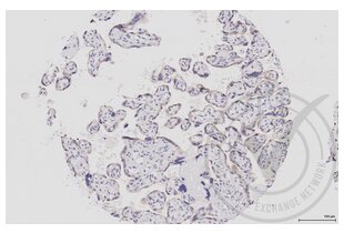
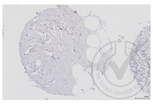
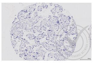
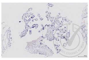
- Immunohistochemistry
- Positive Control
- Human placenta
- Negative Control
- Human breast adipocytes
- Notes
- Signal detected in positive control sample and not in negative control sample.
- Primary Antibody
- Antigen: Mitogen-Activated Protein Kinase Kinase 1 (MAP2K1)
- Catalog number: ABIN686482
- Lot number: 90915
- Secondary Antibody
- Antibody: Biotinylated goat anti-rabbit/anti-mouse (Kit)
- Lot number: D07640BA
- Full Protocol
- Immunohistochemistry was performed on a Ventana NEXes automated platform; instrument manufacturer specific reagents are italicized.
- 1. Slides were preheated in convection oven at 60°C for 30 min
- 2. Deparaffinization procedure: - 3 changes of Xylene, 5 min each - 3 changes of 100% Ethanol, 3 min each - 3 changes of 95% Ethanol, 3 min each - Rinsed in distilled water, 3 changes
- 3. Heat retrieval procedure - Slides retrieved in 10.0 mM Citrate, pH6.0 in a 1000W microwave oven (~100°C) for 15 min. - Slides were allowed to cool (in citrate) for 30 min. - Slides were washed x 3 in Distilled water
- 4. NEXes instrument procedure, iView DAB paraffin protocol (*abridged*): - Slide chamber warmed to 37°C
- 5. Slides rinsed with *reaction buffer* x3
- 6. *iView Inhibitor (H2O2)* applied and incubated for 4 min
- 7. Slides rinsed with *reaction buffer*
- 8. Antibody Application - Primary antibody diluted 1:250 in PBS (100 microliter applied/slide) - Ventana Isotype control applied neat - Slides Incubated overnight at room temperature (~12 hours ~25°C)
- 9. Slides rinsed with *reaction buffer* x3
- 10. *iView Biotinylated IgG* applied and incubated for 8 min
- 11. Slides rinsed with *reaction buffer*
- 14. *iView Streptavidin-Horseradish Peroxidase* applied and incubated for 8 min
- 15. Slides rinsed with *reaction buffer*
- 16. *iView DAB/H2O2* applied and incubated for 8 min
- 17. Slides rinsed with *reaction buffer*
- 18. *iView Copper* applied and incubated for 4 min
- 19. Slides rinsed with *reaction buffer*
- 20. Slides washed in Dawn Detergent/tap water
- 21. Counterstain Procedure - Hematoxylin (Leica 560 MX) 30 sec - Slides washed in tap water, 1 min - Decolorized (10% Acetic Acid in 70% ethanol), 1 min - Slides washed in tap water, 1 min - Bluing (Austin Clear Ammonia), 1 min - Slides washed in tap water, 1 min
- 22. Dehydration/coverslipping procedure: - 3 changes of 95% Ethanol, 3 min each - 3 changes of 100% Ethanol, 3 min each - 3 changes of Xylene, 5 min each - Mounted with Permount
- 23. Imaging: Leica SCN 400F Whole Slide Scanner with Digital Image Hub and Leica Slidepath software
- Experimental Notes
- Deviations from protocol/procedure supplied by manufacturer:
- Step 1: Heated tissue 60°C for 30 minutes; manufacturer heats for 45 minutes.
- Step 2: No ethanol wash was performed during deparaffinization; manufacturer includes 1 wash of 80% ethanol for 3 minutes.
- Step 3.1: Slides were heated for 15 minutes; manufacturer provides a range of 15-20 minutes.
- Step 3.2: Slides were cooled for 30 minutes; manufacturer cools for 20 minutes.
- Step 4: Italicized reagents and incubation time are fixed instrument parameters.
- Step 5: Secondary species-specific serum block not used; manufacturer blocks with 5% normal goat serum for 2 hours.
- Step 8.1: Antibody diluted in PBS at 1:250; manufacture did not recommend diluent or dilution.
- Step 8.2.1: Primary antibody incubated at room temperature overnight; manufacturer incubates overnight 4°C with agitation.
Validation #029646 (Immunohistochemistry)![Successfully validated 'Independent Validation' Badge]()
![Successfully validated 'Independent Validation' Badge]() Validation ImagesFull Methods
Validation ImagesFull Methods -
- Format
- Liquid
- Concentration
- 1 μg/μL
- Buffer
- 0.01M TBS( pH 7.4) with 1 % BSA, 0.02 % Proclin300 and 50 % Glycerol.
- Preservative
- ProClin
- Precaution of Use
- This product contains ProClin: a POISONOUS AND HAZARDOUS SUBSTANCE, which should be handled by trained staff only.
- Storage
- 4 °C,-20 °C
- Storage Comment
- Shipped at 4°C. Store at -20°C for one year. Avoid repeated freeze/thaw cycles.
- Expiry Date
- 12 months
-
- Target
- MEK1 (MAP2K1) (Mitogen-Activated Protein Kinase Kinase 1 (MAP2K1))
- Alternative Name
- MEK1 (MAP2K1 Products)
- Background
-
Synonyms: CFC3, MEK1, MKK1, MAPKK1, PRKMK1, Dual specificity mitogen-activated protein kinase kinase 1, MAP kinase kinase 1, MAPKK 1, ERK activator kinase 1, MAPK/ERK kinase 1, MEK 1, MAP2K1
Background: Dual specificity protein kinase which acts as an essential component of the MAP kinase signal transduction pathway. Binding of extracellular ligands such as growth factors, cytokines and hormones to their cell-surface receptors activates RAS and this initiates RAF1 activation. RAF1 then further activates the dual-specificity protein kinases MAP2K1/MEK1 and MAP2K2/MEK2. Both MAP2K1/MEK1 and MAP2K2/MEK2 function specifically in the MAPK/ERK cascade, and catalyze the concomitant phosphorylation of a threonine and a tyrosine residue in a Thr-Glu-Tyr sequence located in the extracellular signal-regulated kinases MAPK3/ERK1 and MAPK1/ERK2, leading to their activation and further transduction of the signal within the MAPK/ERK cascade. Depending on the cellular context, this pathway mediates diverse biological functions such as cell growth, adhesion, survival and differentiation, predominantly through the regulation of transcription, metabolism and cytoskeletal rearrangements. One target of the MAPK/ERK cascade is peroxisome proliferator-activated receptor gamma (PPARG), a nuclear receptor that promotes differentiation and apoptosis. MAP2K1/MEK1 has been shown to export PPARG from the nucleus. The MAPK/ERK cascade is also involved in the regulation of endosomal dynamics, including lysosome processing and endosome cycling through the perinuclear recycling compartment (PNRC), as well as in the fragmentation of the Golgi apparatus during mitosis.
- Gene ID
- 5604
- UniProt
- Q02750
- Pathways
- MAPK Signaling, RTK Signaling, Interferon-gamma Pathway, Fc-epsilon Receptor Signaling Pathway, Neurotrophin Signaling Pathway, Activation of Innate immune Response, Toll-Like Receptors Cascades, Autophagy, Signaling of Hepatocyte Growth Factor Receptor, BCR Signaling
-


 (1 validation)


