BAD antibody (AA 101-204)
-
- Key Features
-
- High quality BAD Primary Antibody for the detection of BAD.
- Reliable product with high standard validation data
- Target See all BAD Antibodies
- BAD (BCL2-Associated Agonist of Cell Death (BAD))
-
Binding Specificity
- AA 101-204
-
Reactivity
- Human, Mouse, Rat
-
Host
- Rabbit
-
Clonality
- Polyclonal
-
Conjugate
- This BAD antibody is un-conjugated
-
Application
- Western Blotting (WB), ELISA, Immunohistochemistry (Paraffin-embedded Sections) (IHC (p)), Immunofluorescence (Paraffin-embedded Sections) (IF (p)), Immunofluorescence (Cultured Cells) (IF (cc)), Flow Cytometry (FACS), Immunocytochemistry (ICC), Immunohistochemistry (Frozen Sections) (IHC (fro))
- Cross-Reactivity
- Human, Mouse, Rat
- Purification
- Purified by Protein A.
- Immunogen
- KLH conjugated synthetic peptide derived from human BAD
- Isotype
- IgG
- Product Specific Information
-
- What can the BAD Antibody ABIN674709 be used for?
- This polyclonal BAD Antibody detects BAD. The BAD Antibody has been validated for various applications and can be used for the detection of BAD and derivatives by Western Blotting, ELISA, Immunohistochemistry (Paraffin-embedded Sections), Immunofluorescence (Paraffin-embedded Sections), Immunofluorescence (Cultured Cells), Flow Cytometry, Immunocytochemistry, Immunohistochemistry (Frozen Sections).
- What validation data is available for this BAD Antibody?
- The primary antibody has currently 3 product images that show its performance in a variety of applications. The product is currently available in 100 μL quantity. BAD Antibody for the detection of BAD and derivatives.
- What is the function of BAD?
- The protein encoded by this gene is a member of the BCL-2 family. BCL-2 family members are known to be regulators of programmed cell death. This protein positively regulates cell apoptosis by forming heterodimers with BCL-xL and BCL-2, and reversing their death repressor activity. Proapoptotic activity of this protein is regulated through its phosphorylation. Protein kinases AKT and MAP kinase, as well as protein phosphatase calcineurin were found to be involved in the regulation of this protein. Alternative splicing of this gene results in two transcript variants which encode the same isoform. [provided by RefSeq, Jul 2008].
-
-
- Application Notes
-
WB 1:300-5000
ELISA 1:500-1000
FCM 1:20-100
IHC-P 1:200-400
IHC-F 1:100-500
IF(IHC-P) 1:50-200
IF(IHC-F) 1:50-200
IF(ICC) 1:50-200
ICC 1:100-500 - Restrictions
- For Research Use only
-
- by
- Alamo Laboratories Inc
- No.
- #029802
- Date
- 08/26/2014
- Antigen
- Lot Number
- 090915
- Method validated
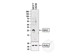
- Western Blotting
- Positive Control
- MCF7 cells
- Negative Control
- C6/36 cells (non-reactive species)
- Notes
- A strong band was observed in the positive control sample at the correct molecular weight. No bands were observed in the negative control sample.
- Primary Antibody
- Antigen: BCL2-Associated Agonist of Cell Death (BAD)
- Catalog number: ABIN674709
- Lot number: 090915
- Antibody Dilution: 1:200
- Secondary Antibody
- Antigen: Goat Anti-Rabbit IgG (H + L)-HRP Conjugate
- Lot number: L170-6515
- Antibody Dilution: 1:10,000
- Full Protocol
- 1. The cell extracts were heated at 95°C for 5 minutes in 1X SDS Sample Buffer containing 1% SDS and 1.25% β-mercaptoethanol.
- 2. 15 μl of heated extracts were loaded and resolved on 8-16% SDS-polyacrylamide gel.
- 3. The Thermo Scientific - Spectra Multicolor Broad Range (Cat # 26634) were used as molecular mass markers.
- 4. Proteins were then transferred onto PVDF membrane by wet transfer and protein transfer was confirmed with Ponceau-S staining.
- 5. The PVDF membrane was incubated with 25 ml of blocking buffer [Tris Buffered Saline, pH 7.4 plus 0.1% TW20 (TBST)] containing 5% (W/V) BSA at room temperature for 1 hour.
- 6. The membrane was rinsed with TBST once.
- 7. The membrane was immersed with the protein side up in the primary antibody solution in TBST containing 5% (W/V) BSA and incubated for 16 hours at 4°C.
- 8. The membrane was rinsed in TBST thrice for 5 minutes each.
- 9. The membrane was incubated in the HRP-conjugated secondary antibody solution in TBST containing 5% (W/V) BSA and incubated for 1 hour at room temperature (~26°C) with gentle agitation.
- 10. The membrane was rinsed thrice TBST thrice for 5 minutes each.
- 11. The membrane was rinsed in TBS twice for 30 seconds each.
- 12. Signals were detected with ECL-2 Substrate. The blot was scanned for 600 seconds.
- 13. The membrane was rinsed three times TBST.
- 14. Incubated in Acidic Glycine Stripping Buffer at room temperature with gentle agitation for 3 times, 10 minutes each.
- 15. The membrane was washed in TBST 2 times for 10 minutes each.
- 16. Repeated Steps 5-12 with the loading control antibody (for Anti-actin) and its matching secondary antibody.
- Experimental Notes
- - No experimental challenges noted.
Validation #029802 (Western Blotting)![Successfully validated 'Independent Validation' Badge]()
![Successfully validated 'Independent Validation' Badge]() Validation ImagesFull Methods
Validation ImagesFull Methods -
- Format
- Liquid
- Concentration
- 1 μg/μL
- Buffer
- 0.01M TBS( pH 7.4) with 1 % BSA, 0.02 % Proclin300 and 50 % Glycerol.
- Preservative
- ProClin
- Precaution of Use
- This product contains ProClin: a POISONOUS AND HAZARDOUS SUBSTANCE, which should be handled by trained staff only.
- Storage
- 4 °C,-20 °C
- Storage Comment
- Shipped at 4°C. Store at -20°C for one year. Avoid repeated freeze/thaw cycles.
- Expiry Date
- 12 months
-
- Target
- BAD (BCL2-Associated Agonist of Cell Death (BAD))
- Alternative Name
- Bad (BAD Products)
- Background
-
Synonyms: BBC2, BCL2L8, Bcl2-associated agonist of cell death, BAD, Bcl-2-binding component 6, Bcl-2-like protein 8, Bcl2-L-8, Bcl-xL/Bcl-2-associated death promoter, Bcl2 antagonist of cell death, BBC6
Background: Promotes cell death. Successfully competes for the binding to Bcl-X(L), Bcl-2 and Bcl-W, thereby affecting the level of heterodimerization of these proteins with BAX. Can reverse the death repressor activity of Bcl-X(L), but not that of Bcl-2 (By similarity). Appears to act as a link between growth factor receptor signaling and the apoptotic pathways.
- Gene ID
- 572
- UniProt
- Q92934
- Pathways
- MAPK Signaling, PI3K-Akt Signaling, RTK Signaling, Apoptosis, Fc-epsilon Receptor Signaling Pathway, Positive Regulation of Peptide Hormone Secretion, Carbohydrate Homeostasis, Positive Regulation of Endopeptidase Activity, Regulation of Carbohydrate Metabolic Process, Hepatitis C, CXCR4-mediated Signaling Events
-


 (1 validation)


