MERTK antibody (AA 501-600)
 (1 validation)
-
- Target See all MERTK Antibodies
- MERTK (C-Mer Proto-Oncogene Tyrosine Kinase (MERTK))
-
Binding Specificity
- AA 501-600
-
Reactivity
- Mouse
-
Host
- Rabbit
-
Clonality
- Polyclonal
-
Conjugate
- This MERTK antibody is un-conjugated
-
Application
- ELISA, Immunohistochemistry (Paraffin-embedded Sections) (IHC (p))
- Cross-Reactivity
- Mouse
- Predicted Reactivity
- Human,Rat
- Purification
- Purified by Protein A.
- Immunogen
- KLH conjugated synthetic peptide derived from human of human MERTK
- Isotype
- IgG
- Top Product
- Discover our top product MERTK Primary Antibody
-
anti-C-Mer Proto-Oncogene Tyrosine Kinase (MERTK) (AA 587-858) antibody
MERTK Reactivity: Human WB, IHC, ICC, IP Host: Rabbit Polyclonal unconjugated
anti-C-Mer Proto-Oncogene Tyrosine Kinase (MERTK) antibodyMERTK Reactivity: Human WB, ELISA, IHC, IF, FACS Host: Rabbit Polyclonal unconjugated
anti-C-Mer Proto-Oncogene Tyrosine Kinase (MERTK) antibodyMERTK Reactivity: Human WB, IF, FACS, IHC (p) Host: Rabbit Polyclonal RB17701 unconjugated
anti-C-Mer Proto-Oncogene Tyrosine Kinase (MERTK) (AA 55-148) antibodyMERTK Reactivity: Human IHC, IHC (f) Host: Mouse Monoclonal MERTK-3015 unconjugated
anti-C-Mer Proto-Oncogene Tyrosine Kinase (MERTK) (AA 402-416), (Extracellular), (N-Term) antibodyMERTK Reactivity: Human, Mouse, Rat WB, IHC, IF, FACS Host: Rabbit Polyclonal unconjugated
anti-C-Mer Proto-Oncogene Tyrosine Kinase (MERTK) (AA 840-999) antibodyMERTK Reactivity: Human WB, ELISA, IHC Host: Rabbit Polyclonal unconjugated
anti-C-Mer Proto-Oncogene Tyrosine Kinase (MERTK) (AA 21-120) antibodyMERTK Reactivity: Human WB, ELISA Host: Mouse Monoclonal 2D2 unconjugated
anti-C-Mer Proto-Oncogene Tyrosine Kinase (MERTK) antibodyMERTK Reactivity: Human ELISA Host: Mouse Monoclonal 7E5G1 unconjugated
anti-C-Mer Proto-Oncogene Tyrosine Kinase (MERTK) (AA 946-980), (C-Term) antibodyMERTK Reactivity: Mouse, Rat WB Host: Rabbit Polyclonal RB50416 unconjugated
anti-C-Mer Proto-Oncogene Tyrosine Kinase (MERTK) (Extracellular Domain) antibodyMERTK Reactivity: Human WB Host: Mouse Monoclonal A311F9G3 unconjugated
-
- Application Notes
-
ELISA 1:500-1000
IHC-P 1:200-400 - Restrictions
- For Research Use only
-
- by
- Palczewski Lab, Center For Translational Vision Research, UC Irvine
- No.
- #104528
- Date
- 12/13/2023
- Antigen
- MERTK
- Lot Number
- AI08084045
- Method validated
- Immunohistochemistry
- Positive Control
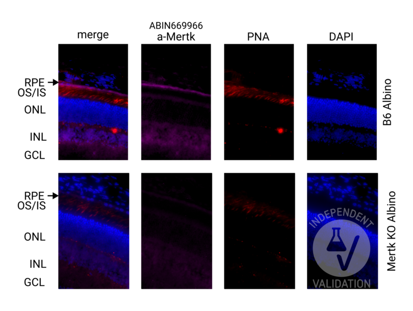
Retina cryosection from B6 Albino (B6(Cg)-Tyrc-2J/J) animal
- Negative Control
Retina cryosection from Mertk-ko Albino animal (B6(Cg)-Tyrc-2J/J background).
- Notes
Passed. Presence of specific signal in the RPE cell layer was considered as indication of specific immunoreactivity.
- Primary Antibody
- ABIN669966
- Secondary Antibody
- anti-rabbit secondary antibody Alexa Fluor 647 (Thermo Fisher Scientific A32795)
- Full Protocol
- Collect eyes from mice and fix with paraformaldehyde 4% (Electron Microscopy Sciences, 15710) in 1x PBS for 30 min at RT
- Cryoprotection with sucrose series:
- Wash in 10% sucrose in 1x PBS
- Immerse in 10% sucrose in 1x PBS for 30 min at RT
- Wash in 20% sucrose in 1x PBS
- Immerse in 20% sucrose in 1x PBS for 30 min RT
- Wash in 30% sucrose in 1x PBS
- 30% sucrose ON at 4°C
- Embed eyes in OCT compound (Tissue-Tek O.C.T. Compound, 4583)
- Cut retinal sections at a thickness of 12 μm on a cryostat
- Air dry sections for 15 min at RT, store at -80°C until use
- Bring sections to RT and rehydrate in 1x PBS for 1 h
- Incubate sections in blocking buffer (1x PBS, 3% BSA (Sigma-Aldrich, A7030), 3% Donkey serum (Sigma-Aldrich, S30-100ML), 0.1% Triton X-100 (Sigma-Aldrich, X100-500ML)) for 1 h at RT
- Incubate sections with primary rabbit anti-MERTK antibody diluted 1:100 in blocking buffer ON at RT. Include MERTK knock-out or no primary antibody negative controls
- Rinse sections 3 times with 1x PBS, 0.1% Triton X100. Keep negative controls in a separate container
- Incubate sections with AF647-conjugated donkey anti-rabbit antibody (Thermo Fisher, A32795, 1:500) and Peanut Agglutinin (PNA) with Rhodamine (Vector Laboratories, RL-1072-5, 1:250) diluted in blocking buffer for 2 h at RT
- Rinse sections once with 1x PBS, 0.1% Triton X-100 for 5 min at RT
- Mount sections in VECTASHIELD® HardSet™ Antifade Mounting Medium with DAPI (Vector Laboratories, H-1500)
- Acquire images with a fluorescence microscope and appropriate filter settings. For the validation purposes Keyence BZ-X800E fluorescence microscope was used with following filters: BZ-X DAPI for DAPI, BZ-X TRITC for Rhodamine, BZ-X Cy5 for AF647. Images were taken at 40x magnification
- Experimental Notes
Experiment involved validation of the specificity of anti-MERTK antibody. MERTK protein is important for eye function and highly expressed specifically in the RPE. Validation is based on comparison of staining performed on wild-type and MERTK-ko cryosections of the mouse retina.
To aid orientation in the cell layers PNA counterstain was included to visualize cone inner and outer segments (IS/OS) that are located adjacent to the RPE.
Validation #104528 (Immunohistochemistry)
 Validation Images
![Retinal sections from the wild-type (B6 albino) and Mertk knock-out (albino) mice immunostained with anti-MERTK antibody ABIN669966. DAPI staining shows localization of the inner (INL) and outer (ONL) nuclear layer of the mouse retina. PNA co-staining was used to visualize cone inner and outer segments (IS/OS) in the retina. Presence of specific signal in the RPE cell layer confirms specific immunoreactivity.]() Retinal sections from the wild-type (B6 albino) and Mertk knock-out (albino) mice immunostained with anti-MERTK antibody ABIN669966. DAPI staining shows localization of the inner (INL) and outer (ONL) nuclear layer of the mouse retina. PNA co-staining was used to visualize cone inner and outer segments (IS/OS) in the retina. Presence of specific signal in the RPE cell layer confirms specific immunoreactivity.
Full Methods
Retinal sections from the wild-type (B6 albino) and Mertk knock-out (albino) mice immunostained with anti-MERTK antibody ABIN669966. DAPI staining shows localization of the inner (INL) and outer (ONL) nuclear layer of the mouse retina. PNA co-staining was used to visualize cone inner and outer segments (IS/OS) in the retina. Presence of specific signal in the RPE cell layer confirms specific immunoreactivity.
Full Methods -
- Format
- Liquid
- Concentration
- 1 μg/μL
- Buffer
- 0.01M TBS( pH 7.4) with 1 % BSA, 0.02 % Proclin300 and 50 % Glycerol.
- Preservative
- ProClin
- Precaution of Use
- This product contains ProClin: a POISONOUS AND HAZARDOUS SUBSTANCE, which should be handled by trained staff only.
- Storage
- 4 °C,-20 °C
- Storage Comment
- Shipped at 4°C. Store at -20°C for one year. Avoid repeated freeze/thaw cycles.
- Expiry Date
- 12 months
-
- Target
- MERTK (C-Mer Proto-Oncogene Tyrosine Kinase (MERTK))
- Alternative Name
- MERTK (MERTK Products)
- Background
-
Synonyms: MER, RP38, c-Eyk, c-mer, Tyro12, Tyrosine-protein kinase Mer, Proto-oncogene c-Mer, Receptor tyrosine kinase MerTK, MERTK
Background: Receptor tyrosine kinase that transduces signals from the extracellular matrix into the cytoplasm by binding to several ligands including LGALS3, TUB, TULP1 or GAS6. Regulates many physiological processes including cell survival, migration, differentiation, and phagocytosis of apoptotic cells (efferocytosis). Ligand binding at the cell surface induces autophosphorylation of MERTK on its intracellular domain that provides docking sites for downstream signaling molecules. Following activation by ligand, interacts with GRB2 or PLCG2 and induces phosphorylation of MAPK1, MAPK2, FAK/PTK2 or RAC1. MERTK signaling plays a role in various processes such as macrophage clearance of apoptotic cells, platelet aggregation, cytoskeleton reorganization and engulfment. Functions in the retinal pigment epithelium (RPE) as a regulator of rod outer segments fragments phagocytosis. Plays also an important role in inhibition of Toll-like receptors (TLRs)-mediated innate immune response by activating STAT1, which selectively induces production of suppressors of cytokine signaling SOCS1 and SOCS3.
- Gene ID
- 10461
- UniProt
- Q12866
- Pathways
- RTK Signaling
-


