STAT5A antibody
 (1 validation)
-
- Target See all STAT5A Antibodies
- STAT5A (Signal Transducer and Activator of Transcription 5A (STAT5A))
-
Reactivity
- Human, Mouse, Rat
-
Host
- Rabbit
-
Clonality
- Monoclonal
-
Conjugate
- This STAT5A antibody is un-conjugated
-
Application
- Western Blotting (WB), Immunohistochemistry (Paraffin-embedded Sections) (IHC (p)), Immunofluorescence (Cultured Cells) (IF (cc))
- Cross-Reactivity
- Human, Mouse, Rat
- Purification
- Purified by Protein A.
- Immunogen
- Recombinant human STAT5a protein, around C-terminal 100aa.
- Clone
- 6E5
- Isotype
- IgG
-
anti-Signal Transducer and Activator of Transcription 5A (STAT5A) (AA 583-794) antibody
STAT5A Reactivity: Human WB, IHC, ELISA, FACS, ICC Host: Mouse Monoclonal 6D4 unconjugated
anti-Signal Transducer and Activator of Transcription 5A (STAT5A) (AA 1-794) antibodySTAT5A Reactivity: Human WB, IF Host: Mouse Polyclonal unconjugated
anti-Signal Transducer and Activator of Transcription 5A (STAT5A) (AA 1-104) antibodySTAT5A Reactivity: Human WB, ELISA, PLA Host: Mouse Monoclonal 1B12 unconjugated
anti-Signal Transducer and Activator of Transcription 5A (STAT5A) (AA 635-794) antibodySTAT5A Reactivity: Human WB, IF Host: Rabbit Polyclonal unconjugated
anti-Signal Transducer and Activator of Transcription 5A (STAT5A) (pSer780) antibodySTAT5A Reactivity: Human, Mouse, Rat WB, IHC, ELISA, IF, IP, ICC Host: Rabbit Polyclonal unconjugated
anti-Signal Transducer and Activator of Transcription 5A (STAT5A) (pTyr694) antibodySTAT5A Reactivity: Human, Mouse, Rat WB, IHC, ELISA, IF, ICC Host: Rabbit Polyclonal unconjugated
anti-Signal Transducer and Activator of Transcription 5A (STAT5A) (C-Term) antibodySTAT5A Reactivity: Human, Mouse, Rat WB, IHC, ELISA, IF, ICC Host: Rabbit Polyclonal unconjugated
anti-Signal Transducer and Activator of Transcription 5A (STAT5A) (pTyr694) antibodySTAT5A Reactivity: Human WB, IF, IHC (p) Host: Rabbit Polyclonal RB5956 unconjugated
anti-Signal Transducer and Activator of Transcription 5A (STAT5A) (AA 61-160) antibodySTAT5A Reactivity: Human, Mouse, Rat, Cow WB, ELISA, IHC (p), IF (cc), IF (p), IHC (fro) Host: Rabbit Polyclonal unconjugated
anti-Signal Transducer and Activator of Transcription 5A (STAT5A) (AA 763-797), (C-Term) antibodySTAT5A Reactivity: Human, Mouse WB, IHC (p) Host: Rabbit Polyclonal RB52392 unconjugated
-
- Application Notes
-
WB 1:300-5000
IHC-P 1:200-400
IF(ICC) 1:50-200 - Restrictions
- For Research Use only
-
- by
- Gianluca Zambanini, Anna Nordin and Claudio Cantù; Cantù Lab, Gene Regulation during Development and Disease, Linköping University
- No.
- #104384
- Date
- 12/07/2022
- Antigen
- STAT5A
- Lot Number
- BA7271967
- Method validated
- Cleavage Under Targets and Release Using Nuclease
- Positive Control
Polyclonal rabbit anti-H3K4me (antibodies-online, ABIN3023251)
- Negative Control
Polyclonal guinea pig anti-rabbit IgG (antibodies-online, ABIN101961)
- Notes
Passed. ABIN5557532 allows for CUT&RUN targeting of STAT5A in mouse forelimb tissue.
- Primary Antibody
- ABIN5557532
- Secondary Antibody
- Full Protocol
- Cell harvest and nuclear extraction
- Dissect 3 Fore limbs (11.5 DAC) from mouse strain RjOrl:SWISS for each sample.
- Dissociate the tissue into single cells in TrypLE (Thermo Fisher Scientific) for 15 min at 37 °C.
- Centrifuge cell solution 5 min at 800 x g at RT.
- Remove the liquid carefully.
- Gently resuspend cells in 1 mL of Nuclear Extraction Buffer (20 mM HEPES-KOH pH 8.2, 20% Glycerol, 0.05% IGEPAL, 0.5 mM Spermidine, 10 mM KCl, Roche Complete Protease Inhibitor EDTA-free).
- Move the solution to a 2 mL centrifuge tube.
- Pellet the nuclei 800 x g for 5 min.
- Repeat the NE wash twice for a total of three washes.
- Resuspend the nuclei in 20 µL NE Buffer per sample.
- Concanavalin A beads preparation
- Prepare one 2 mL microcentrifuge tube.
- Gently resuspend the magnetic Concanavalin A Beads (antibodies-online, ABIN6952467).
- Pipette 20 µL Con A Beads slurry for each sample into the 2 mL microcentrifuge tube.
- Place the tube on a magnet stand until the fluid is clear. Remove the liquid carefully.
- Remove the microcentrifuge tube from the magnetic stand.
- Pipette 1 mL Binding Buffer (20 mM HEPES pH 7.5, 10 mM KCl, 1 mM CaCl2, 1 mM MnCl2) into the tube and resuspend ConA beads by gentle pipetting.
- Spin down the liquid from the lid with a quick pulse in a table-top centrifuge.
- Place the tubes on a magnet stand until the fluid is clear. Remove the liquid carefully.
- Remove the microcentrifuge tube from the magnetic stand.
- Repeat the wash twice for a total of three washes.
- Gently resuspend the ConA Beads in a volume of Binding Buffer corresponding to the original volume of bead slurry, i.e. 20 µL per sample.
- Nuclei immobilization – binding to Concanavalin A beads
- Carefully vortex the nuclei suspension and add 20 µL of the Con A beads in Binding Buffer to the cell suspension for each sample.
- Close tube tightly incubates 10 min at 4 °C.
- Put the 2 mL tube on the magnet stand and when the liquid is clear remove the supernatant.
- Resuspend the beads in 1 mL of EDTA Wash buffer (20 mM HEPES pH 7.5, 150 mM NaCl, 0.5 mM Spermidine, Roche Complete Protease Inhibitor EDTA-free, 2mM EDTA).
- Incubate 5 min at RT.
- Place the tube on the magnet stand and when the liquid is clear remove the supernatant.
- Resuspend the beads in 200 µl of Wash Buffer (20 mM HEPES pH 7.5, 150 mM NaCl, 0.5 mM Spermidine, Roche Complete Protease Inhibitor EDTA-free) per sample.
- Primary antibody binding
- Divide nuclei suspension into separate 200 µL PCR tubes, one for each antibody (150,000 cells per sample).
- Add 2 µL antibody (anti-STAT5A antibody ABIN5557532, anti-H3K4me positive control antibody ABIN3023251, and guinea pig anti-rabbit IgG negative control antibody ABIN101961) to the respective tube, corresponding to a 1:100 dilution.
- Incubate at 4 °C ON.
- Place the tubes on a magnet stand until the fluid is clear. Remove the liquid carefully.
- Remove the microcentrifuge tubes from the magnetic stand.
- Wash with 200 µL of Wash Buffer using a multichannel pipette to accelerate the process.
- Repeat the wash five times for a total of six washes.
- pAG-MNase Binding
- Prepare a 1.5 mL microcentrifuge tube containing 100 µL of pAG mix per sample (100 µL of wash buffer + 58.5 µg pAG-MNase per sample).
- Place the PCR tubes with the sample on a magnet stand until the fluid is clear. Remove the liquid carefully.
- Remove tubes from the magnetic stand.
- Resuspend the beads in 100 µL of pAG-MNase premix.
- Incubate 30 min at 4 °C.
- Place the tubes on a magnet stand until the fluid is clear. Remove the liquid carefully.
- Remove the microcentrifuge tubes from the magnetic stand.
- Wash with 200 µL of Wash Buffer using a multichannel pipette to accelerate the process.
- Repeat the wash five times for a total of six washes.
- Resuspend in 100 µL of Wash Buffer.
- MNase digestion and release of pAG-MNase-antibody-chromatin complexes
- Place PCR tubes on ice and allow to chill.
- Prepare a 1.5 mL microcentrifuge tube with 102 µl of 2 mM CaCl2 mix per sample (100 µl Wash Buffer + 2 µL 100 mM CaCl2) and let it chill on ice.
- Always in ice, place the samples on the magnetic rack and when the liquid is clear remove the supernatant.
- Resuspend the samples in 100 µl of the 2 mM CaCl2 mix and incubate in ice for exactly 30 min.
- Place the sample on the magnet stand and when the liquid is clear remove the supernatant.
- Resuspend the sample in 50 µl of 1x Urea STOP Buffer (8.5 M Urea, 100 mM NaCl, 2 mM EGTA, 2 mM EDTA, 0.5% IGEPAL).
- Incubate the samples 1h at 4°C.
- Transfer the supernatant containing the pAG-MNase-bound digested chromatin fragments to fresh 200 µl PCR tubes.
- DNA Clean up
- Take the Mag-Bind® TotalPure NGS beads (Omega Bio-Tek, M1378-01) from the storage and wait until they are at RT.
- Add 2x volume of beads to each sample (e.g. 100 µL of beads for 50 µL of sample).
- Incubate the beads and the sample for 15 min at RT.
- During incubation prepare fresh EtOH 80%.
- Place the PCR tubes on a magnet stand and when the liquid is clear remove the supernatant.
- Add 200 µl of fresh 80% EtOH to the sample without disturbing the beads (Important!!! Do NOT resuspend the beads or remove the tubes from the magnet stand or the sample will be lost).
- Incubate 30 sec at RT.
- Remove the EtOH from the sample.
- Repeat the wash with 80% EtOH.
- Resuspend the beads in 25 µL of 10 mM Tris-HCl pH 8.2.
- Incubate the sample for 2 min at RT.
- Repeat the 2x beads clean up as described before (this time with 50 µL of beads for each sample).
- Resuspend the beads + DNA in 20 µL of 10 mM Tris-HCl pH 8.2.
- Library preparation and sequencing
- Prepare Libraries using KAPA HyperPrep Kit using KAPA Dual-Indexed adapters according to protocol.
- Sequence samples on an Illumina NextSeq 500 sequencer, using a NextSeq 500/550 High Output Kit v2.5 (75 Cycles), 36 bp PE.
- Peak calling
- Trim reads using using bbTools bbduk (BBMap - Bushnell B. - sourceforge.net/projects/bbmap/) to remove adapters, artifacts and repeat sequences.
- Map aligned reads to the hg38 human genome using bowtie with options -m 1 -v 0 -I 0 -X 500.
- Use SAMtools to convert SAM files to BAM files and remove duplicates.
- Use BEDtools genomecov to produce Bedgraph files.
- Call peaks using SEACR with a 0.001 threshold and the option norm stringent.
- Experimental Notes
The protocol is published in Zambanini, G. et al. A New CUT&RUN Low Volume-Urea (LoV-U) protocol uncovers Wnt/β-catenin tissue-specific genomic targets. Development (2022). PMID 36355069
Validation #104384 (Cleavage Under Targets and Release Using Nuclease)
 Validation Images
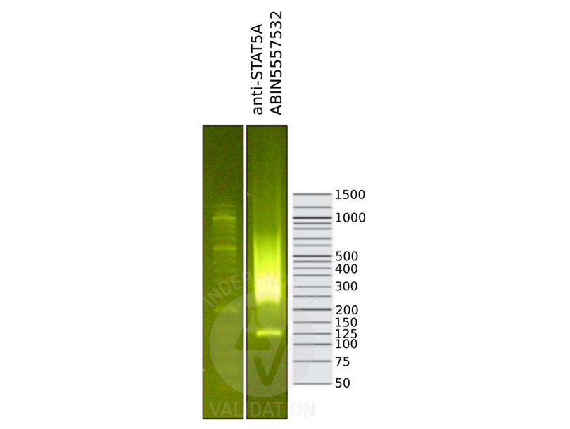
![Library profiles comparing fragment size distributions on an E-Gel EX 2% agarose gel (Thermo Fisher). Fragments obtained from CUT&RUN using STAT5A ABIN5557532 (right) after library preparation, compared to the E-Gel Sizing DNA Ladder (Thermo Fisher) (left).]() Library profiles comparing fragment size distributions on an E-Gel EX 2% agarose gel (Thermo Fisher). Fragments obtained from CUT&RUN using STAT5A ABIN5557532 (right) after library preparation, compared to the E-Gel Sizing DNA Ladder (Thermo Fisher) (left).
Library profiles comparing fragment size distributions on an E-Gel EX 2% agarose gel (Thermo Fisher). Fragments obtained from CUT&RUN using STAT5A ABIN5557532 (right) after library preparation, compared to the E-Gel Sizing DNA Ladder (Thermo Fisher) (left).
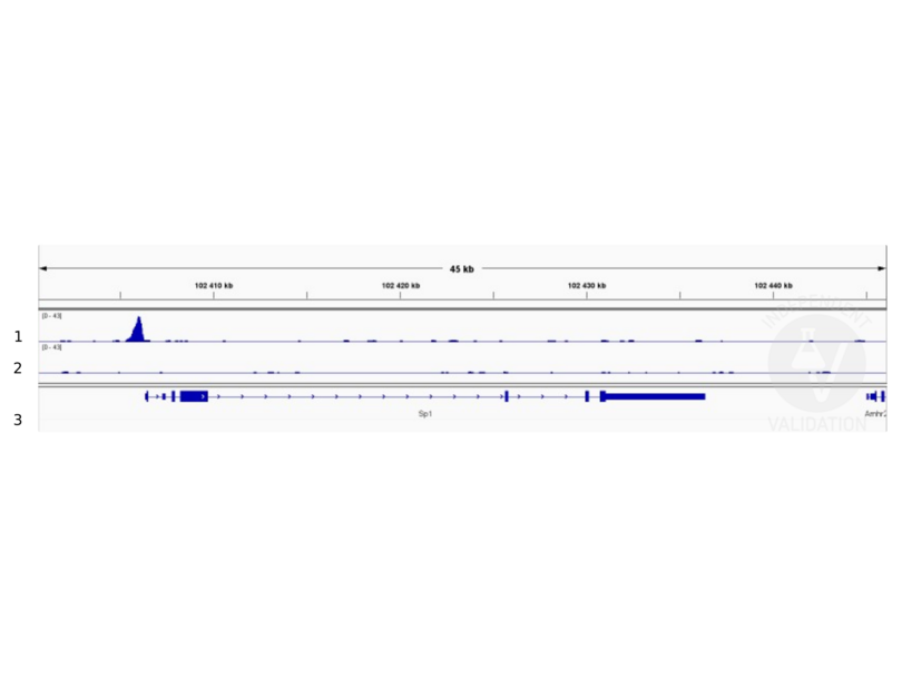
![1. Alignment tracks from CUT&RUN targeting STAT5A in mouse fore limb (11.5) cells using ABIN5557532, showing the Sp1 locus. 2. Alignment tracks using negative control IgG, ABIN1019613. 3. RefSeq Genes.]() 1. Alignment tracks from CUT&RUN targeting STAT5A in mouse fore limb (11.5) cells using ABIN5557532, showing the Sp1 locus. 2. Alignment tracks using negative control IgG, ABIN1019613. 3. RefSeq Genes.
Full Methods
1. Alignment tracks from CUT&RUN targeting STAT5A in mouse fore limb (11.5) cells using ABIN5557532, showing the Sp1 locus. 2. Alignment tracks using negative control IgG, ABIN1019613. 3. RefSeq Genes.
Full Methods -
- Format
- Liquid
- Concentration
- 1 μg/μL
- Buffer
- Aqueous buffered solution containing 1xTBS ( pH 7.4), 1 % BSA, 40 %Glycerol and 0.05 % Sodium Azide.
- Preservative
- ProClin
- Precaution of Use
- This product contains ProClin: a POISONOUS AND HAZARDOUS SUBSTANCE, which should be handled by trained staff only.
- Storage
- 4 °C,-20 °C
- Storage Comment
- Shipped at 4°C. Store at -20°C for one year. Avoid repeated freeze/thaw cycles.
- Expiry Date
- 12 months
-
- Target
- STAT5A (Signal Transducer and Activator of Transcription 5A (STAT5A))
- Alternative Name
- STAT5a (STAT5A Products)
- Background
-
Synonyms: MGF, STAT5, Signal transducer and activator of transcription 5A, STAT5A
Background: Carries out a dual function: signal transduction and activation of transcription. Mediates cellular responses to the cytokine KITLG/SCF and other growth factors. Mediates cellular responses to ERBB4. May mediate cellular responses to activated FGFR1, FGFR2, FGFR3 and FGFR4. Binds to the GAS element and activates PRL-induced transcription. Regulates the expression of milk proteins during lactation.
- Gene ID
- 6776
- UniProt
- P42229
- Pathways
- JAK-STAT Signaling, RTK Signaling, Response to Growth Hormone Stimulus, C21-Steroid Hormone Metabolic Process, Regulation of Leukocyte Mediated Immunity, Positive Regulation of Immune Effector Process, CXCR4-mediated Signaling Events, Activated T Cell Proliferation
-


