MFN1 antibody (AA 1-234)
-
- Target See all MFN1 Antibodies
- MFN1 (Mitofusin 1 (MFN1))
-
Binding Specificity
- AA 1-234
-
Reactivity
- Mouse
-
Host
- Rabbit
-
Clonality
- Polyclonal
-
Conjugate
- This MFN1 antibody is un-conjugated
-
Application
- Western Blotting (WB), Immunohistochemistry (IHC), Immunoprecipitation (IP), Immunocytochemistry (ICC)
- Purpose
- Polyclonal Antibody to Mitofusin 1 (MFN1)
- Specificity
- The antibody is a rabbit polyclonal antibody raised against MFN1. It has been selected for its ability to recognize MFN1 in immunohistochemical staining and western blotting.
- Cross-Reactivity
- Human, Rat
- Purification
- Antigen-specific affinity chromatography followed by Protein A affinity chromatography
- Immunogen
- Recombinant Mitofusin 1 (MFN1) corresdonding to Met1~Ile234
- Isotype
- IgG
- Top Product
- Discover our top product MFN1 Primary Antibody
-
-
- Application Notes
-
Western blotting: 0.5-2 μg/mL
1:500-2000 Immunohistochemistry: 5-20 μg/mL
1:50-200 Immunocytochemistry: 5-20 μg/mL
1:50-200 Optimal working dilutions must be determined by end user.
- Comment
-
The thermal stability is described by the loss rate. The loss rate was determined by accelerated thermal degradation test, that is, incubate the protein at 37°C for 48h, and no obvious degradation and precipitation were observed. The loss rate is less than 5% within the expiration date under appropriate storage condition.
- Restrictions
- For Research Use only
-
- by
- Wang Lab, Department of Animal Science, College of Agriculture & Natural Resources
- No.
- #103013
- Date
- 05/30/2018
- Antigen
- MFN1
- Lot Number
- A20180411903
- Method validated
- Western Blotting
- Positive Control
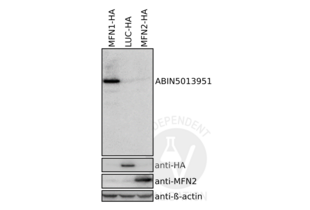
293T cells transfected with MFN1-HA expression vector; the full length MFN1 CDS has been inserted upstream of an HA-tag in plasmid pcDNA3.1.
- Negative Control
293T cells transfected with an LUC-HA expression vector, and the full length LUC CDS has been inserted upstream of an HA-tag in plasmid pcDNA3.1.
293T cells transfected with MFN2-HA expression vector; the full length MFN2 CDS has been inserted upstream of an FLAG-tag in plasmid pcDNA3.1.
- Notes
Passed. ABIN5013951 specifically recognizes antigen in sample.
- Primary Antibody
- ABIN5013951
- Secondary Antibody
- goat anti-rabbit IgG (H+L) antibody HRP conjugate (Bio-Rad, 170-6515)
- Full Protocol
- Grow 293T cells in DMEM medium (Gibco, lot 1897400) supplemented with 10% fetal bovine serum (Gibco, lot 1913756) and 1% Penicillin-Streptomycin (thermo, 10378016), at 37°C and 7% CO2 in 6-well plates to 60-70% confluency.
- Transfect cells with plasmids expressing either MFN1-HA, MFN2-HA or LUC-HA with max 2µg of plasmid using PEI transfection reagent (Poly-Science, 24765-1) following the manufacturer´s instructions.
- Lyse cells in 100µl RIPA buffer containing protease inhibitors (Sigma-Aldrich, P8340).
- Determine total protein content of the lysates using a Bradford protein assay (Protein Assay Dye Reagent Concentrate, Bio-Rad, 5000006).
- Denature 100µg total protein for 10min at 95°C in 1x SDS loading buffer and subsequently separate them on a freshly cast 10% denaturing polyacrylamide gel.
- Transfer proteins onto Immun-Blot Low Fluorescence PVDF Membrane (0.45μm) at 1.0A and up to 25V for 30min via Trans-Blot Turbo Transfer System.
- Block the membrane with Bio-rad Blotting-Grade Blocker (5% non-fat milk in PBS).
- Incubation with primary
- rabbit anti-MFN1 antibody (antibodies-online, ABIN5013951, lot A20180411903) diluted 1:200 in Blocking Buffer (5% non-fat milk) ON at 4°C.
- mouse anti-HA-tag antibody (Santa Cruz, sc-7392) diluted 1:2000 in Blocking Buffer (5% non-fat milk) ON at 4°C (only LUC-HA expressing cells).
- mouse anti-MFN2-tag antibody (Santa Cruz, sc-100560) diluted 1:2000 in Blocking Buffer (5% non-fat milk) ON at 4°C.
- mouse anti-beta-actin antibody (Sigma-Aldrich, A1978) diluted 1:10000 in Blocking Buffer(5% non-fat milk) ON at 4°C.
- Wash membrane 3x for 10min with PBS supplemented with 0.1% Tween 20.
- Incubation with secondary goat anti-rabbit IgG (H+L) antibody HRP conjugate (Bio-Rad, 170-6515) or goat anti-mouse IgG (H+L) antibody HRP conjugate (Bio-Rad, 170-6516) diluted 1:10000 in Odyssey Blocking Buffer for 1h at RT.
- Wash membrane 3x for 10min with PBS supplemented with 0.1% Tween 20.
- Add Clarity and Clarity Max Western ECL Blotting Substrates and scan the membrane on a Bio-Rad ChemiDoc XRS+System.
- Experimental Notes
The antigen antibody ABIN5013951 reveals a protein of the expected molecular weight of antigen in lysates of cells. The protein bands is only visible in the positive but not the negative controls.
Validation #103013 (Western Blotting)![Successfully validated 'Independent Validation' Badge]()
![Successfully validated 'Independent Validation' Badge]() Validation ImagesFull Methods
Validation ImagesFull Methods -
- Format
- Liquid
- Concentration
- Lot specific
- Buffer
- 0.01M PBS, pH 7.4, containing 0.05 % Proclin-300, 50 % glycerol.
- Preservative
- ProClin
- Storage
- 4 °C,-20 °C
- Storage Comment
- Store at 4°C for frequent use. Stored at -20°C in a manual defrost freezer for two year without detectable loss of activity. Avoid repeated freeze-thaw cycles.
- Expiry Date
- 24 months
-
- Target
- MFN1 (Mitofusin 1 (MFN1))
- Alternative Name
- Mitofusin 1 (MFN1 Products)
- Background
- Hfzo1, Hfzo2, Transmembrane GTPase MFN1, Fzo homolog
-


 (1 validation)


