TDC2 antibody (C-Term)
Quick Overview for TDC2 antibody (C-Term) (ABIN4889606)
Target
Reactivity
Host
Clonality
Application
-
-
Binding Specificity
- C-Term
-
Specificity
- Reacts with Drosophila melanogaster 72 kDa Tdc2 protein
-
Purification
- Purified (protein A)
-
Immunogen
- Synthetic peptide derived from C-terminal part of Drosophila Tdc2 protein.
-
-
-
Application Notes
-
Working dilution: Optimal dilutions should be determined by the end user.
The following are guidelines only:
IHC(1:200 - 1:1000) WB(1:200 - 1:2000) -
Restrictions
- For Research Use only
-
-
- by
- Department of Entomology, University of California, Riverside
- No.
- #100812
- Date
- 05/26/2017
- Antigen
- Tdc2
- Lot Number
- 13B1
- Method validated
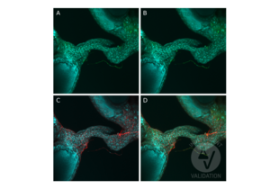
- Immunofluorescence
- Positive Control
D. melanogaster octopaminergic neurons labeled with Tdc2-Gal4 of the abdominal nerve to the ovary
- Negative Control
- Notes
Passed. ABIN809182 labels octopaminergic neurons specifically and with no background.
- Primary Antibody
- ABIN4889606
- Secondary Antibody
- goat anti-rabbit AF542 conjugated antibody (Life Technologies)
- Full Protocol
- Dissect ovaries of D. melanogaster ETHR-Gal4/UAS-MCD8-GFP expressing GFP in octopaminergic neurons in cold Schneider’s Insect Medium (S2; Sigma Aldrich, S01416).
- Transfer tissue to 2ml protein LoBind tubes (Eppendorf, 022431102) filled with S2 containing 2% paraformaldehyde (PFA) at RT.
- Fix tissue for 55min at RT while nutating.
- Wash tissue 4x 10min with 1.75ml PBS containing 0.5% Triton X-100 (PBST).
- Remove PBST and add 200µl 5% goat serum (GS; Thermo Fisher Scientific, 16210064) in PBST per tube.
- Incubate 1.5h at RT on a rotator.
- Remove blocking solution.
- Incubate with primary
- rabbit anti-Tdc2 antibody (Tyrosine Decarboxylase 2) (C-Term) (antibodies-online, ABIN4889606, lot 13B1) diluted 1:200 in blocking solution.
- mouse anti-GFP (Thermo Fisher Scientific) diluted 1:500 in blocking solution.
- Incubate for 4h at RT followed by 36-48h at 4°C on a rotator.
- Rinse tissue with 1.75ml PBST. Allow the tissue to settle to the bottom before removing the liquid.
- Wash tissue 3x 30min with 1.75ml PBST.
- Incubate with secondary 200µl secondary goat anti-rabbit AF542 conjugated antibody (Life Technologies) and goat anti-mouse AF488 conjugated antibody (Life Technologies, A11034) diluted 1:500 in blocking solution containing 0.5mg/ml DAPI.
- Incubate for 4h at RT followed by 72h at 4°C on a rotator.
- Rinse tissue with 1.75ml PBST. Allow the tissue to settle to the bottom before removing the liquid.
- Wash tissue 3x 30min with 1.75ml PBST.
- Add 1.75ml PBST containing 4% PFA at RT.
- Fix tissue for 5h at RT while nutating.
- Rinse tissue with 1.75ml PBST. Allow the tissue to settle to the bottom before removing the liquid.
- Wash tissue 4x 15min with 1.75ml PBST.
- Mount tissue on a poly-L-lysine (Sigma Aldrich, P1524-25MG) coated cover glass.
- Dehydrate tissue by covering the cover glass for 10min each with 30%, 50%, 75%, 95%, 100%, 100%, and 100% EtOH.
- Clearing using by covering the cover glass 3x 5min with xylene.
- Add 7 drops of dibutyl phthalate in xylene (DPX) on top of the mounted tissue.
- Seat cover glass face down gently onto a prepared slide with spacers.
- Let the slide dry for 48h at RT in a hood before viewing.
- Experimental Notes
Staining of ETHR-Gal4/UAS-MCD8-GFP expressing GFP in octopaminergic neurons was performed as previously described.
ABIN4889606 worked fantastically. It labeled octopaminergic neurons specifically and with no background. Pictures are below. The well-characterized neurons labeled with Tdc2-Gal4 of the abdominal nerve to the ovary described in Middleton et al. (2006) overlapped with ABIN4889606. Staining with ABIN4889606 was stronger than Gal4 labeling.
Validation #100812 (Immunofluorescence)![Successfully validated 'Independent Validation' Badge]()
![Successfully validated 'Independent Validation' Badge]() Validation ImagesFull Methods
Validation ImagesFull Methods -
-
Format
- Lyophilized
-
Reconstitution
- Must be reconstituted in distilled water.
-
Concentration
- 1 mg/mL
-
Buffer
- Tris 0,1M, glycine 0,1M, sucrose 2 %
-
Storage
- 4 °C/-20 °C
-
Storage Comment
- Lyophilized powder stable for a minimum of 2 years at -20°C. Store reconstituted antibodies at +4°C. For extended periods store in aliquots at -20°C. Antibodies are guaranteed for 6 month from date of receipt.
-
Expiry Date
- 24 months
-
-
- TDC2 (Tyrosine Decarboxylase 2 (TDC2))
-
Alternative Name
- Tyrosine Decarboxylase 2
-
Background
- Enzyme involved in tyrosine metabolism. Use Pyridoxal phosphate as cofactor.
-
Gene ID
- 246620
-
UniProt
- A1Z6N4
Target
-


 (1 validation)


