SLC22A6 antibody (C-Term)
-
- Target See all SLC22A6 Antibodies
- SLC22A6 (Solute Carrier Family 22 Member 6 (SLC22A6))
-
Binding Specificity
- AA 534-550, C-Term
-
Reactivity
- Human, Rat
-
Host
- Rabbit
-
Clonality
- Polyclonal
-
Conjugate
- This SLC22A6 antibody is un-conjugated
-
Application
- Western Blotting (WB), Immunohistochemistry (Paraffin-embedded Sections) (IHC (p))
- Purpose
- Rabbit IgG polyclonal antibody for Solute carrier family 22 member 6(SLC22A6) detection. Tested with WB, IHC-P in Human,Rat.
- Sequence
- QKYMVPLQAS AQEKNGL
- Cross-Reactivity (Details)
- No cross reactivity with other proteins.
- Characteristics
-
Rabbit IgG polyclonal antibody for Solute carrier family 22 member 6(SLC22A6) detection. Tested with WB, IHC-P in Human,Rat.
Gene Name: solute carrier family 22(organic anion transporter), member 6
Protein Name: Solute carrier family 22 member 6 - Purification
- Immunogen affinity purified.
- Immunogen
- A synthetic peptide corresponding to a sequence at the C-terminus of human SLC22A6(534-550aa QKYMVPLQASAQEKNGL), different from the related rat sequence by three amino acids.
- Isotype
- IgG
-
-
- Application Notes
-
WB: Concentration: 0.1-0.5 μg/mL, Tested Species: Human
IHC-P: Concentration: 0.5-1 μg/mL, Tested Species: Human, Rat, Epitope Retrieval by Heat: Boiling the paraffin sections in 10 mM citrate buffer, pH 6.0, for 20 mins is required for the staining of formalin/paraffin sections.
Notes: Tested Species: Species with positive results. Predicted Species: Species predicted to be fit for the product based on sequence similarities. Other applications have not been tested. Optimal dilutions should be determined by end users. - Comment
-
Antibody can be supported by chemiluminescence kit ABIN921124 in WB, supported by ABIN921231 in IHC(P).
- Restrictions
- For Research Use only
-
- by
- Human Protein Atlas
- No.
- #300029
- Date
- 12/13/2016
- Antigen
- SLC22A6
- Lot Number
- Method validated
- Immunohistochemistry
- Positive Control
- kidney
- Negative Control
- Notes
- Passed. SLC22A6 staining with ABIN3044261 is consistent with the literature.
- Primary Antibody
- ABIN3044261
- Secondary Antibody
- HRP polymer (ThermoFisher Scientific, TL-125-PH)
- Full Protocol
- Sample preparation:
- Fixation and TMA preparation according to PMID: 22688270.
- Cut 4µm TMA sections using a waterfall microtome (ThermoFisher Scientific, HM355S).
- Dry sections ON at RT and then back them for 12-24h at 50°C.
- Deparaffinization and rehydration in xylene and graded alcohol series:
- xylene twice for 3min.
- 100% ethanol for 1min.
- 95% ethanol for 1min.
- 0.3% H2O2 in 95% ethanol for 5min to block endogenous peroxidase.
- 70% ethanol for 1min.
- 50% ethanol for 1min.
- distilled H2O for 2min.
- Antigen Retrieval by Heat Induced Epitope Retrieval (HIER):
- Boil the sections in Lab Vision Citrate buffer pH6.0 (ThermoFisher Scientific, TA-250-PM1X) for 4min at 125°C in a decloaking chamber (Biocare Medical).
- Allow slides to cool down to 90°C in the decloaking chamber.
- Immunostaining in a Lab Vision Autostainer 480 (ThermorFisher Scientific) at RT with 300µl of reagents for each step:
- Rinse sections in Lab Vision wash buffer with extra tween added to a final concentration of 0.2% (ThermoFisher Scientific, TA-999-TT and TA-125-TW) wash buffer.
- Block sections with Lab Vision Ultra V-Block (ThermoFisher Scientific, TA-125-UB) for 5min.
- Rinse sections 2x in wash buffer.
- Incubate sections with primary rabbit anti-SLC22A6 antibody (antibodies-online, ABIN3044261) diluted 1:1100 for 30min.
- Rinse sections 3x in wash buffer.
- Incubate sections with labeled HRP polymer (ThermoFisher Scientific, TL-125-PH) for 30min.
- Rinse sections 2x in wash buffer.
- Develop in DAB Quanto solution (ThermoFisher Scientific, TL-125-QHDX) for 5min.
- Rinse sections in distilled H2O.
- Washing, counterstaining, and coverslipping in Autostainer XL (Leica Biosystems, ST5010) at RT:
- Counterstaining in Mayer’s hematoxylin plus (HistoLab, 01820) for 7.5min.
- Rinse sections in tap water for 5min.
- Rinse sections in lithium carbonate water diluted 1:5 for 5min.
- Dehydration in graded ethanol series and Neo-Clear (Merck Millipore, 109843) according to the manufacturer recommendations.
- Automated mounting (Leica Biosystems, CV5030) of coverslip with Pertex (Histolab, 00871).
- Microscopy:
- Image acquisition on a Leica Aperio AT2 and Aperio Scanscope AT at 20x magnification.
- Experimental Notes
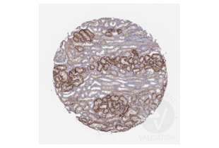
- Based on published literature, SLC22A6 is expected to be expressed in the kidney. ABIN3044261 shows basal staining in renal tubules. Additional staining is observed in a subset of immune cells but at a lower level.
Validation #300029 (Immunohistochemistry)![Successfully validated 'Independent Validation' Badge]()
![Successfully validated 'Independent Validation' Badge]() Validation ImagesFull Methods
Validation ImagesFull Methods -
- Format
- Lyophilized
- Reconstitution
- Add 0.2 mL of distilled water will yield a concentration of 500 μg/mL.
- Concentration
- 500 μg/mL
- Buffer
- Each vial contains 5 mg BSA, 0.9 mg NaCl, 0.2 mg Na2HPO4, 0.05 mg Thimerosal, 0.05 mg Sodium azide.
- Preservative
- Thimerosal (Merthiolate), Sodium azide
- Precaution of Use
- This product contains Sodium azide and Thimerosal (Merthiolate): POISONOUS AND HAZARDOUS SUBSTANCES which should be handled by trained staff only.
- Handling Advice
- Avoid repeated freezing and thawing.
- Storage
- 4 °C/-20 °C
- Storage Comment
-
At -20°C for one year. After reconstitution, at 4°C for one month.
It can also be aliquotted and stored frozen at -20 °C for a longer time. Avoid repeated freezing and thawing. - Expiry Date
- 12 months
-
- Target
- SLC22A6 (Solute Carrier Family 22 Member 6 (SLC22A6))
- Alternative Name
- SLC22A6 (SLC22A6 Products)
- Background
-
SLC22A6(Solute carrier family 22(organic anion transporter), member 6), also called OAT1 or PAHT, is a protein that in humans is encoded by the SLC22A6 gene, which is also a member of the organic anion transporter(OAT) family of proteins. OAT1 is a transmembrane protein that is expressed in the brain, the placenta, the eyes, smooth muscles, and the basolateral membrane of proximal tubular cells of the kidneys. The SLC22A6 gene is mapped on 11q12.3. It plays a central role in renal organic anion transport. Along with OAT3, OAT1 mediates the uptake of a wide range of relatively small and hydrophilic organic anions from plasma into the cytoplasm of the proximal tubular cells of the kidneys. The SLC22A6 gene contains 10 exons and spans 8.2 kb. OAT1 functions as organic anion exchanger. When the uptake of one molecule of an organic anion is transported into a cell by an OAT1 exchanger, one molecule of an endogenous dicarboxylic acid(such as glutarate, ketoglutarate, etc) is simultaneously transported out of the cell. PAH uptake in Xenopus oocytes injected with OAT1 mRNA was demonstrated by Race et al.
Synonyms: FLJ55736 antibody|hOAT1 antibody|hPAHT antibody|hROAT1 antibody|MGC45260 antibody|OAT1 antibody|Organic anion transporter 1 antibody|OTTHUMP00000236796 antibody|OTTHUMP00000236797 antibody|OTTHUMP00000236798 antibody|OTTHUMP00000236799 antibody|PAH transporter antibody|PAHT antibody|Para aminohippurate transporter antibody|Renal organic anion transporter 1 antibody|ROAT1 antibody|S22A6_HUMAN antibody|SLC22A6 antibody|Solute carrier family 22(organic anion transporter) member 6 antibody|Solute carrier family 22 member 6 antibody - UniProt
- Q4U2R8
- Pathways
- Dicarboxylic Acid Transport
-


 (1 validation)


