MAP2 antibody
-
- Target See all MAP2 Antibodies
- MAP2 (Microtubule-Associated Protein 2 (MAP2))
-
Reactivity
- Human, Mouse, Pig
-
Host
- Mouse
-
Clonality
- Monoclonal
-
Conjugate
- This MAP2 antibody is un-conjugated
-
Application
- Western Blotting (WB), ELISA, Immunocytochemistry (ICC), Immunoprecipitation (IP), Immunohistochemistry (Paraffin-embedded Sections) (IHC (p)), Immunohistochemistry (Frozen Sections) (IHC (fro))
- Specificity
- The antibody MT-08 recognizes an epitope (aa 1375-1395) located in central domain of molecule Microtubule Associated Protein 2ab (MAP2ab), an intracellular antigen.
- Cross-Reactivity (Details)
- Human, Porcine, Mouse
- Purification
- Purified by protein-A affinity chromatography.
- Purity
- > 95 % (by SDS-PAGE)
- Immunogen
- Microtubule protein (bovine brain) enriched for kinesin
- Clone
- MT-08
- Isotype
- IgG1
-
-
- Application Notes
-
Immunohistochemistry (paraffin sections): Recommended dilution: 10 μg/mL, positive tissue: brain.
Immunohistochemistry (frozen sections): Positive tissue: murine brain.
Immunoprecipitation: Positive material: porcine brain.
Western blotting: Positive control: porcine brain.
ELISA: Positive control: porcine brain.
Immunocytochemistry: Positive control: human neuroblastoma SH-SY5Y. - Restrictions
- For Research Use only
-
- by
- Akoya Biosciences
- No.
- #104333
- Date
- 04/20/2021
- Antigen
- MAP2
- Lot Number
- 536958
- Method validated
- Multiplex Immunohistochemistry
- Positive Control
Fresh frozen mouse olfactory bulb
- Negative Control
Unlabeled control (mouse fresh frozen)
- Notes
Passed. The anti-MAP2 antibody ABIN125739 specifically labels structural elements of neurons. Staining can be observed in neuronal cell bodies, axons and occasionally in proximal dendritic processes.
- Primary Antibody
- ABIN125739
- Secondary Antibody
- Full Protocol
- Protocol details are described in the Akoya Biosciences CODEX® User Manual (see https://www.akoyabio.com/wp-content/uploads/2021/01/CODEX-User-Manual.pdf).
- Tissue preparation as outlined in the Akoya Biosciences CODEX® User Manual fresh-frozen tissue protocol.
- Conjugation of the anti-MAP2 antibody ABIN125739 to an oligo barcode used to bind oligo-bound fluorophore ATTO 550.
- Experimental Notes
No signal was detected in unlabeled specimens.
Specific staining of MAP2 was also observed with human FFPE cortical tissue sections with both citrate antigen retrieval and EDTA antigen retrieval.
Optimal staining and signal to noise ratios were obtained if tissue was pre-treated for autofluorescence removal (see https://www.akoyabio.com/wp-content/uploads/2020/07/Customer-Demonstrated-Protocol-Autofluorescence-Quenching-Mar2020.pdf).
Validation #104333 (Multiplex Immunohistochemistry)![Successfully validated 'Independent Validation' Badge]()
![Successfully validated 'Independent Validation' Badge]() Validation Images
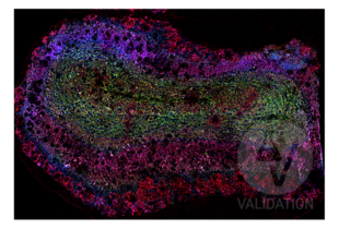
Validation Images![Murine fresh frozen coronal olfactory bulb section (Thickness = 5 µm) labeled with anti-MAP2 antibody ABIN125739 (blue; bound to fluorophore ATTO 550). Labeling is present throughout olfactory bulb layers with and concentration in the external plexiform and mitral cell layers. Slc17a7 and PSD-95/DLG4 were labeled with ABIN1027710 (green; bound to fluorophore ATTO 550) and ABIN361694 (red; bound to fluorophore ATTO 550).]() Murine fresh frozen coronal olfactory bulb section (Thickness = 5 µm) labeled with anti-MAP2 antibody ABIN125739 (blue; bound to fluorophore ATTO 550). Labeling is present throughout olfactory bulb layers with and concentration in the external plexiform and mitral cell layers. Slc17a7 and PSD-95/DLG4 were labeled with ABIN1027710 (green; bound to fluorophore ATTO 550) and ABIN361694 (red; bound to fluorophore ATTO 550).
Murine fresh frozen coronal olfactory bulb section (Thickness = 5 µm) labeled with anti-MAP2 antibody ABIN125739 (blue; bound to fluorophore ATTO 550). Labeling is present throughout olfactory bulb layers with and concentration in the external plexiform and mitral cell layers. Slc17a7 and PSD-95/DLG4 were labeled with ABIN1027710 (green; bound to fluorophore ATTO 550) and ABIN361694 (red; bound to fluorophore ATTO 550).
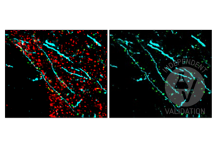
![FFPE normal human cortex tissue section labeled with anti-MAP2 antibody ABIN125739 (cyan; bound to fluorophore ATTO 550) after EDTA antigen retrieval. DLG4 and Synapsin were labeled with anti-DLG4 antibody ABIN361694 (green; bound to fluorophore ATTO 550) and anti-SYN1 antibody ABIN5542390 (red; bound to fluorophore AF488).]() FFPE normal human cortex tissue section labeled with anti-MAP2 antibody ABIN125739 (cyan; bound to fluorophore ATTO 550) after EDTA antigen retrieval. DLG4 and Synapsin were labeled with anti-DLG4 antibody ABIN361694 (green; bound to fluorophore ATTO 550) and anti-SYN1 antibody ABIN5542390 (red; bound to fluorophore AF488).
Full Methods
FFPE normal human cortex tissue section labeled with anti-MAP2 antibody ABIN125739 (cyan; bound to fluorophore ATTO 550) after EDTA antigen retrieval. DLG4 and Synapsin were labeled with anti-DLG4 antibody ABIN361694 (green; bound to fluorophore ATTO 550) and anti-SYN1 antibody ABIN5542390 (red; bound to fluorophore AF488).
Full Methods -
- Concentration
- 1 mg/mL
- Buffer
- Phosphate buffered saline (PBS), pH 7.4, 15 mM sodium azide
- Preservative
- Sodium azide
- Precaution of Use
- This product contains Sodium azide: a POISONOUS AND HAZARDOUS SUBSTANCE which should be handled by trained staff only.
- Handling Advice
- Do not freeze.
- Storage
- 4 °C
- Storage Comment
- Store at 2-8°C. Do not freeze.
-
-
: "Specific nuclear localizing sequence directs two myosin isoforms to the cell nucleus in calmodulin-sensitive manner." in: PLoS ONE, Vol. 7, Issue 1, pp. e30529, (2012) (PubMed).
-
: "Specific nuclear localizing sequence directs two myosin isoforms to the cell nucleus in calmodulin-sensitive manner." in: PLoS ONE, Vol. 7, Issue 1, pp. e30529, (2012) (PubMed).
-
- Target
- MAP2 (Microtubule-Associated Protein 2 (MAP2))
- Alternative Name
- MAP2ab (MAP2 Products)
- Background
- Microtubule associated protein 2,MAP2a and 2b (270 kDa) being found mostly in dendrites, stabilize microtubules (shift the reaction kinetics in addition of new subunits and microtubule growth) and participate in determining the structure of different parts of vertebrate nerve cells.,Microtubule-associated protein 2, MAP-2
- Gene ID
- 4133
- UniProt
- P11137
-


 (1 reference)
 (1 validation)


