beta Catenin antibody (C-Term)
-
- Target See all beta Catenin (CATNB) Antibodies
- beta Catenin (CATNB) (Catenin, beta (CATNB))
-
Binding Specificity
- C-Term
-
Reactivity
- Human, Zebrafish (Danio rerio)
-
Host
- Rabbit
-
Clonality
- Polyclonal
-
Conjugate
- This beta Catenin antibody is un-conjugated
-
Application
- Western Blotting (WB), Immunohistochemistry (IHC), ELISA
- Supplier Product No.
- 600-401-c68
- Supplier
- Rockland
- Purpose
- beta Catenin Antibody
- Cross-Reactivity (Details)
- beta catenin antibody is directed against catenin beta-1 protein.
- Characteristics
- Synonyms: rabbit anti-beta Catenin Antibody, rabbit anti-Catenin beta antibody, catenin beta-1, ß-catenin-1, ß-catenin, CTNNB1, CTNNB, beta-catenin antibody
- Purification
- The product was affinity purified from monospecific antiserum by immunoaffinity chromatography.
- Sterility
- Sterile filtered
- Immunogen
-
Immunogen: beta catenin antibody was prepared from whole rabbit serum produced by repeated immunizations with a synthetic peptide corresponding to catenin beta-1 C-terminus.
Immunogen Type: Conjugated Peptide
- Isotype
- IgG
- Top Product
- Discover our top product CATNB Primary Antibody
-
-
- Application Notes
-
Immunohistochemistry Dilution: 5 - 10 μg/mL
Application Note: beta catenin antibody has been tested for use in ELISA, western blotting, immunohistochemistry, and ISH. Specific conditions for reactivity should be optimized by the end user. Expect a band approximately 85.5 kDa in size corresponding to catenin beta-1 protein by western blotting in the appropriate cell lysate or extract.
Western Blot Dilution: 1:500
ELISA Dilution: 1:75,000 - 1:125,000
Other: ISH
- Restrictions
- For Research Use only
-
- by
- ADS Biosystems Inc
- No.
- #029816
- Date
- 09/18/2014
- Antigen
- Lot Number
- 27397
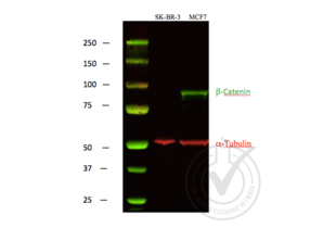
- Method validated
- Western Blotting
- Positive Control
- MCF-7 cells
- Negative Control
- SK-BR-3 cells
- Notes
- A strong specific band was observed in the positive control at the expected size (~85.5 kDa) that is not observed in the negative control.
- Primary Antibody
- Antigen: Beta Catenin
- Catalog number: ABIN1043907
- Lot number: 27397
- Dilution: 1:1,000
- Secondary Antibody
- Antibody: IRDye 680LT Goat Anti-Rabbit
- Lot number: C30725-01
- Dilution: 1:10,000
- Full Protocol
- Lysates were mixed with NuPAGE® LDS Sample Buffer (Life Technologies NP0007) and NuPAGE® Sample Reducing Agent (Life Technologies NP0004) and denatured for 5 minutes at 90ºC.
- 40 μg of each lysate was electrophoresed on a Bolt 4-12% Bis-Tris Gel (Life Technologies BG04120BOX) and run in Bolt MOPS SDS Running Buffer (Life Technologies B0001) at 160 volts for 1 hour.
- Odyssey Western Protein Standard (LI-COR #928-40000) was run as a molecular weight standard.
- PVDF membrane was activated with methanol.
- Protein samples were transferred to activated PVDF membrane in a wet Bolt Transfer Apparatus (Life Technologies B1000) at room temperature for 1 hour at 20 volts (started at 230mA, ended at 110mA).
- The membrane was blocked in x LI-COR Odyssey WB block solution for 1 hour at room temperature.
- The membrane was incubated with the primary antibody diluted 1:1000 in x LI-COR Odyssey WB block solution incubated 2 hours at room temperature.
- The membrane was washed 4 x 5 minutes in 1 x PBS-T (PBS solution with 0.1% Tween 20).
- The membrane was incubated with IRDye® 800CW Goat anti-Mouse Secondary Antibody (Red) and IRDye 680LT Goat Anti-Rabbit Secondary Antibody (Green) from LI-COR (#827-11081, Lot #C30725-01), both 1:10,000 dilutions. Incubation was performed at room temperature for 45 minutes.
- The membrane was washed 4 x 5 minutes in 1 x PBS-T (PBS solution with 0.1% Tween 20).
- Proteins were detected using Odyssey machine scanning with green channel for loading control and red channel for potential LPL band.
- Experimental Notes
- - No experimental challenges noted.
Validation #029816 (Western Blotting)![Successfully validated 'Independent Validation' Badge]()
![Successfully validated 'Independent Validation' Badge]() Validation ImagesFull Methods
Validation ImagesFull Methods -
- Format
- Liquid
- Concentration
- 1.15 mg/mL
- Buffer
-
Buffer: 0.02 M Potassium Phosphate, 0.15 M Sodium Chloride, pH 7.2
Stabilizer: None
Preservative: 0.01 % (w/v) Sodium Azide - Preservative
- Sodium azide
- Precaution of Use
- This product contains Sodium azide: a POISONOUS AND HAZARDOUS SUBSTANCE which should be handled by trained staff only.
- Storage
- 4 °C,-20 °C
- Storage Comment
- Store vial at -20° C prior to opening. Aliquot contents and freeze at -20° C or below for extended storage. Avoid cycles of freezing and thawing. Centrifuge product if not completely clear after standing at room temperature. This product is stable for several weeks at 4° C as an undiluted liquid. Dilute only prior to immediate use.
- Expiry Date
- 12 months
-
-
: "Wnt status-dependent oncogenic role of BCL9 and BCL9L in hepatocellular carcinoma." in: Hepatology international, Vol. 14, Issue 3, pp. 373-384, (2020) (PubMed).
: "The terminal region of beta-catenin promotes stability by shielding the Armadillo repeats from the axin-scaffold destruction complex." in: The Journal of biological chemistry, Vol. 284, Issue 41, pp. 28222-31, (2009) (PubMed).
-
: "Wnt status-dependent oncogenic role of BCL9 and BCL9L in hepatocellular carcinoma." in: Hepatology international, Vol. 14, Issue 3, pp. 373-384, (2020) (PubMed).
-
- Target
- beta Catenin (CATNB) (Catenin, beta (CATNB))
- Alternative Name
- beta Catenin (CATNB Products)
- Background
- Background: Beta-catenin 1 (or ß-catenin 1) is a protein that is encoded by the CTNNB1 gene. ß-catenin 1 is a subunit of the cadherin protein complex and has been implicated as an integral component in the Wnt signaling pathway. This pathway plays a key role in the regulation of cellular processes involved in development, differentiation, and adult tissue homeostasis. In the presence of Wnt ligand, ß-catenin 1 is not ubiquitinated and accumulates in the nucleus, where it associates with T-cell factor (TCF) family members to regulate target gene expression in many developmental and adult tissues. Recruitment of b-catenin 1 to Wnt response element (WRE) chromatin converts TCFs from transcriptional repressors to activators. ß-catenin 1 is also involved in the regulation of cell adhesion. It acts as a negative regulator of centrosome cohesion. Aberrant Wnt/ß-catenin signaling is widely implicated in cancer, bone disorders, kidney and intestinal cell disorders and other disease states. ß-catenin 1 is located in the cytoplasm when it is unstabilized or bound to CDH1. Interaction with GLIS2 and MUC1 promotes nuclear translocation. Interaction with EMD inhibits nuclear localization. The majority of ß-catenin 1 is localized to the cell membrane. In interphase, colocalizes with CROCC between CEP250 puncta at the proximal end of centrioles, and this localization is dependent on CROCC and CEP250. In mitosis, when NEK2 activity increases, it localizes to centrosomes at spindle poles independent of CROCC. It further co-localizes with CDK5 in the cell-cell contacts and plasma membrane of undifferentiated and differentiated neuroblastoma cells.
- Gene ID
- 1499
- NCBI Accession
- NP_001091679
- UniProt
- F1QGH7
- Pathways
- Peptide Hormone Metabolism
-


 (2 references)
 (1 validation)


