DYKDDDDK Tag antibody
-
- Target See all DYKDDDDK Tag products
- DYKDDDDK Tag
- Reactivity
- Please inquire
-
Host
- Rabbit
-
Clonality
- Polyclonal
-
Conjugate
- This DYKDDDDK Tag antibody is un-conjugated
-
Application
- Western Blotting (WB), ELISA, Immunoprecipitation (IP)
- Supplier Product No.
- 600-401-383
- Supplier
- Rockland
- Sequence
- DYKDDDDK
- Characteristics
- Concentration Definition: by UV absorbance at 280 nm
- Immunogen
- This antibody was purified from whole rabbit serum prepared by repeated immunizations with the Enterokinase (ECS) peptide DYKDDDDK (Asp-Tyr-Lys-Asp-Asp-Asp-Asp-Lys) conjugated to KLH using maleimide. Residues of glycine and cysteine were added to the carboxy terminal end to facilitate coupling. This antibody reacts with DYKDDDDK conjugated proteins.
- Isotype
- IgG
-
-
- Application Notes
- This antibody is optimally suited for monitoring the expression of DYKDDDDK tagged fusion proteins. As such, this antibody can be used to identify fusion proteins containing the DYKDDDDK epitope. The antibody recognizes the epitope tag fused to either the amino- or carboxy- termini of targeted proteins. This antibody has been tested by ELISA and western blotting against both the immunizing peptide and DYKDDDDK containing recombinant proteins. Although not tested, this antibody is likely functional for immunoprecipitation, immunocytochemistry, and other immunodetection techniques. The epitope tag peptide sequence was first derived from the 11-amino-acid leader peptide of the gene-10 product from bacteriophage T7. Now the most commonly used hydrophilic octapeptide is DYKDDDDK. polyclonal antibody to detect DYKDDDDK conjugated proteins binds DYKDDDDK containing fusion proteins with greater affinity than the widely used monoclonal M1, M2 and M5 clones, and shows greater sensitivity in most assays. Affinity purification of the polyclonal antibody results in very low background levels in assays and low cross-reactivity with other cellular proteins.
- Restrictions
- For Research Use only
-
- by
- Group Muehlemann, Department of Chemistry and Biochemistry, University of Bern
- No.
- #101368
- Date
- 05/06/2017
- Antigen
- DYKDDDDK Tag
- Lot Number
- 33451
- Method validated
- Western Blotting
- Positive Control
- HeLa cells expressing DYKDDDDK-tagged THOC1 or THOC5
- Negative Control
- HeLa cells
- Notes
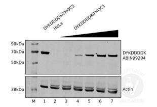
Passed. The DYKDDDDK Tag antibody ABIN99294 specifically recognizes DYKDDDDK-tagged proteins in HeLa cell extracts.
- Primary Antibody
- ABIN99294
- Secondary Antibody
- rabbit anti-actin antibody (Sigma-Aldrich, A5060)
- Full Protocol
- HeLa cells are grown in DMEM/F12 W/L-GLUT (Thermo Fisher Scientific, 32500035) supplemented with 10% FCS (Amimed) and Penicillin-Strepomycin (BioConcept 4-01F00-H), at 37°C and 5% CO2 to 70-80% confluency.
- Transfect cells with an expression plasmids encoding DYKDDDDK-tagged THOC proteins using Lipofectamine 2000 (Thermo Fisher Scientific following the manufacturer´s instructions.
- Grow cells for 48h.
- Trypsinize cells, collect and count them.
- Lyse 107 cells in 1ml cold RIPA buffer (Cold Spring Harbour protocols) containing protease inhibitors (Biotool, B14003).
- Dilute the equivalent of 2x10⁵ cells (20µl) 1:2 with 2x Laemmli SDS sample buffer and heat samples up for 5min to 95°C.
- Separate the denatured samples on a denaturing freshly cast polyacrylamide gel.
- Transfer proteins onto a nitrocellulose membrane (GE Healthcare).
- Block the membrane with TBScontaining 0.5% Tween 20 and 5% milk powder for 30min at RT.
- Incubation with primary antibody
- rabbit anti-DYKDDDDK Tag antibody (antibodies-online, ABIN99294, lot 33451) or
- rabbit anti-actin antibody (Sigma-Aldrich, A5060)
- diluted 1:2000 in TBST 1h at RT or ON at 4°.
- Wash membrane 3x for 10min with TBST.
- Incubation with secondary antibody IRDye 800CW Goat anti-Rabbit (LiCor, 926-32211) diluted 1:10000 in TBST for 1h at RT.
- Wash membrane 3x for 10min with TBST.
- Reveal protein bands on a LI-COR Odyssey imaging system (LI-COR).
- Experimental Notes
ABIN99294 proved a very good antibody for the detection of DYKDDDDK-tagged THOC proteins in Western blot.
The antibody can be reused up to ten times as long as the incubation buffer contains some sodium azide.
The antibody worked in various blocking buffers we tried containing milk or BSA and as well in Pierce WB enhancer.
Validation #101368 (Western Blotting)![Successfully validated 'Independent Validation' Badge]()
![Successfully validated 'Independent Validation' Badge]() Validation Images
Validation Images![Increasing amounts of lysates from HeLa cells transiently expressing DYKDDDDK-tagged THOC1 were separated on an SDS-PAGE and revealed using ABIN99294 as decribed in the protocol section (lanes 3 to 7). Transiently expressed DYKDDDDK-tagged THOC5 can is also be detected using ABIN99294 (lane 1). Untransfected HeLa cell extracts were used as negative controls (lane 2).]() Increasing amounts of lysates from HeLa cells transiently expressing DYKDDDDK-tagged THOC1 were separated on an SDS-PAGE and revealed using ABIN99294 as decribed in the protocol section (lanes 3 to 7). Transiently expressed DYKDDDDK-tagged THOC5 can is also be detected using ABIN99294 (lane 1). Untransfected HeLa cell extracts were used as negative controls (lane 2).
Full Methods
Increasing amounts of lysates from HeLa cells transiently expressing DYKDDDDK-tagged THOC1 were separated on an SDS-PAGE and revealed using ABIN99294 as decribed in the protocol section (lanes 3 to 7). Transiently expressed DYKDDDDK-tagged THOC5 can is also be detected using ABIN99294 (lane 1). Untransfected HeLa cell extracts were used as negative controls (lane 2).
Full Methods -
- by
- Group Muehlemann, Department of Chemistry and Biochemistry, University of Bern
- No.
- #101369
- Date
- 05/06/2017
- Antigen
- DYKDDDDK Tag
- Lot Number
- 33451
- Method validated
- Immunofluorescence
- Positive Control
- HeLa cells expressing DYKDDDDK-tagged EGFP or GAPDH
- Negative Control
- HeLa cells
- Notes
Passed. The DYKDDDDK Tag antibody ABIN99294 specifically labels DYKDDDDK-tagged proteins in HeLa cells in immunofluorescence.
- Primary Antibody
- ABIN99294
- Secondary Antibody
- chicken anti-rabbit AF488 antibody conjugate (Thermo Fisher Scientific, A-21441)
- Full Protocol
- Prepare sterile 10mm cover slips (VWR, 631-1576) in empty 6-well plates.
- Seed 2x10⁵ HeLa cells expressing DYKDDDDK-tagged proteins per well (6-well plate) onto the cover slips one day prior to fixation and let them grow in DMEM/F12 W/L-GLUT (Thermo Fisher Scientific, 32500035) with 10% FCS (Amimed) and antibiotics (Penicillin-Strepomycin 4-01F00-H, Bioconcept) at 37°C and 5% CO2 to 60-80% confluency.
- Wash cells with PBS.
- Remove culture medium and fix the cells with 1.5ml/well 4% PFA for 20-30 min at RT.
- Wash samples 3x 5min with TBS (20mM Tris-HCl, pH 7.5, 150mM NaCl).
- Incubate cells with permeabilization/blocking buffer for 30-60 min at RT.
- Incubate slides with primary rabbit anti-DYKDDDDK Tag antibody (antibodies-online, ABIN99294, lot 33451) diluted 1:200 in 100µl TBS++ (1xTBS, 0.1% Triton X100, 6% serum (or BSA 1.25gr/250mL PBS)) ON at 4°C. A negative control was incubated without primary antibody.
- Take the slides out of the fridge and leave them for 2h at RT.
- Wash slides 3x 5min with TBS++.
- Incubate slides with secondary chicken anti-rabbit AF488 antibody conjugate (Thermo Fisher Scientific, A-21441) for 1.5h at 37°C.
- Incubate slides for 30min at RT.
- Wash slides 2x with TBS.
- Counterstain with 100μL DAPI diluted to 100ng/ml in TBS for 10min at RT.
- Wash slides 3x with TBS.
- Add a drop of homemade Mowiol on the slide and mount the coverslips.
- Cover the borders with nail polish to fix the coverslips to the side.
- Take images on a Leica DMI6000 fluorescence microscope.
- Experimental Notes
Staining of transiently expressed DYKDDDDK-tagged EGFP and GAPDH with ABIN99294 resulted in the expected staining pattern.
Validation #101369 (Immunofluorescence)![Successfully validated 'Independent Validation' Badge]()
![Successfully validated 'Independent Validation' Badge]() Validation Images
Validation Images![HeLa cells transiently expressing DYKDDDDK-tagged EGFP (top row) or GAPDH (middle row) and mock-transfected HeLa cells (bottom row) were stained using DAPI (left column) or DYKDDDDK Tag antibody ABIN99294 in combination with an AF488 conjugated secondary antibody (middle column). The right column shows merged DAPI and ABIN99294 staining.]() HeLa cells transiently expressing DYKDDDDK-tagged EGFP (top row) or GAPDH (middle row) and mock-transfected HeLa cells (bottom row) were stained using DAPI (left column) or DYKDDDDK Tag antibody ABIN99294 in combination with an AF488 conjugated secondary antibody (middle column). The right column shows merged DAPI and ABIN99294 staining.
Full Methods
HeLa cells transiently expressing DYKDDDDK-tagged EGFP (top row) or GAPDH (middle row) and mock-transfected HeLa cells (bottom row) were stained using DAPI (left column) or DYKDDDDK Tag antibody ABIN99294 in combination with an AF488 conjugated secondary antibody (middle column). The right column shows merged DAPI and ABIN99294 staining.
Full Methods -
- Format
- Liquid
- Concentration
- 1.04 mg/mL
- Buffer
- 0.02 M Potassium Phosphate, 0.15 M Sodium Chloride, pH 7.2
- Preservative
- Sodium azide
- Precaution of Use
- This product contains sodium azide: a POISONOUS AND HAZARDOUS SUBSTANCE which should be handled by trained staff only.
- Storage
- -20 °C
-
-
: "LITESEC-T3SS - Light-controlled protein delivery into eukaryotic cells with high spatial and temporal resolution." in: Nature communications, Vol. 11, Issue 1, pp. 2381, (2020) (PubMed).
: "Modulation of virus-induced NF-κB signaling by NEMO coiled coil mimics." in: Nature communications, Vol. 11, Issue 1, pp. 1786, (2020) (PubMed).
: "Pathogenic mutations reveal a role of RECQ4 in mitochondrial RNA:DNA hybrid formation and resolution." in: Scientific reports, Vol. 10, Issue 1, pp. 17033, (2020) (PubMed).
: "Connexin 50 Functions as an Adhesive Molecule and Promotes Lens Cell Differentiation." in: Scientific reports, Vol. 7, Issue 1, pp. 5298, (2019) (PubMed).
: "Construction of a tri-chromatic reporter cell line for the rapid and simple screening of splice-switching oligonucleotides targeting DMD exon 51 using high content screening." in: PLoS ONE, Vol. 13, Issue 5, pp. e0197373, (2018) (PubMed).
: "Resolving the Combinatorial Complexity of Smad Protein Complex Formation and Its Link to Gene Expression." in: Cell systems, Vol. 6, Issue 1, pp. 75-89.e11, (2018) (PubMed).
: "Evolutionary divergence of the sex-determining gene MID uncoupled from the transition to anisogamy in volvocine algae." in: Development (Cambridge, England), Vol. 145, Issue 7, (2018) (PubMed).
: "SARS-Coronavirus Open Reading Frame-3a drives multimodal necrotic cell death." in: Cell death & disease, Vol. 9, Issue 9, pp. 904, (2018) (PubMed).
: "Protease-activated receptor-4 and purinergic receptor P2Y12 dimerize, co-internalize, and activate Akt signaling via endosomal recruitment of β-arrestin." in: The Journal of biological chemistry, Vol. 292, Issue 33, pp. 13867-13878, (2017) (PubMed).
: "Protease-activated Receptor-4 Signaling and Trafficking Is Regulated by the Clathrin Adaptor Protein Complex-2 Independent of β-Arrestins." in: The Journal of biological chemistry, Vol. 291, Issue 35, pp. 18453-64, (2017) (PubMed).
: "Recycling and Endosomal Sorting of Protease-activated Receptor-1 Is Distinctly Regulated by Rab11A and Rab11B Proteins." in: The Journal of biological chemistry, Vol. 291, Issue 5, pp. 2223-36, (2016) (PubMed).
: "S6K-STING interaction regulates cytosolic DNA-mediated activation of the transcription factor IRF3. ..." in: Nature immunology, Vol. 17, Issue 5, pp. 514-522, (2016) (PubMed).
: "GPCR sorting at multivesicular endosomes." in: Methods in cell biology, Vol. 130, pp. 319-32, (2016) (PubMed).
: "Association of FKBP51 with Priming of Autophagy Pathways and Mediation of Antidepressant Treatment Response: Evidence in Cells, Mice, and Humans." in: PLoS medicine, Vol. 11, Issue 11, pp. e1001755, (2014) (PubMed).
: "Essential role of the chaperonin CCT in rod outer segment biogenesis." in: Investigative ophthalmology & visual science, Vol. 55, Issue 6, pp. 3775-85, (2014) (PubMed).
: "Splice isoforms of phosducin-like protein control the expression of heterotrimeric G proteins." in: The Journal of biological chemistry, Vol. 288, Issue 36, pp. 25760-8, (2014) (PubMed).
: "eIF4E-bound mRNPs are substrates for nonsense-mediated mRNA decay in mammalian cells." in: Nature structural & molecular biology, Vol. 20, Issue 6, pp. 710-7, (2013) (PubMed).
: "Metal allergens nickel and cobalt facilitate TLR4 homodimerization independently of MD2." in: EMBO reports, Vol. 13, Issue 12, pp. 1109-15, (2013) (PubMed).
: "Loss of the methyl lysine effector protein PHF20 impacts the expression of genes regulated by the lysine acetyltransferase MOF." in: The Journal of biological chemistry, Vol. 287, Issue 1, pp. 429-37, (2012) (PubMed).
: "Vectors for expression and secretion of FLAG epitope-tagged proteins in mammalian cells." in: BioTechniques, Vol. 20, Issue 1, pp. 136-41, (1996) (PubMed).
-
: "LITESEC-T3SS - Light-controlled protein delivery into eukaryotic cells with high spatial and temporal resolution." in: Nature communications, Vol. 11, Issue 1, pp. 2381, (2020) (PubMed).
-
- Target
- DYKDDDDK Tag
- Abstract
- DYKDDDDK Tag Products
- Target Type
- Tag
- Background
- Epitope tags are short peptide sequences that are easily recognized by tag-specific antibodies. Due to their small size, epitope tags do not affect the biochemical properties of the tagged protein. Most often, sequences encoding the epitope tag are included with the target DNA at the time of cloning to produce fusion proteins containing the epitope tag sequence. This allows Anti epitope tag antibodies to serve as universal detection reagents for any tag-containing protein produced by recombinant means. This means that anti-epitope tag antibodies are a useful alternative to generating specific antibodies to identify, immunoprecipitate or immunoaffinity purify a recombinant protein. The anti-epitope tag antibody is usually functional in a variety of antibody-dependent experimental procedures.
-


 (20 references)
 (2 validations)


