JAG1 antibody (N-Term)
 (2 references)
 (1 validation)
-
- Target See all JAG1 Antibodies
- JAG1 (Jagged 1 (JAG1))
-
Binding Specificity
- N-Term
-
Reactivity
- Human, Mouse
-
Host
- Rabbit
-
Clonality
- Polyclonal
-
Conjugate
- This JAG1 antibody is un-conjugated
-
Application
- Western Blotting (WB), ELISA, Immunohistochemistry (IHC), Immunofluorescence (IF), Fluorescence Microscopy (FM)
- Purpose
- Jagged 1 Antibody
- Cross-Reactivity (Details)
- This protein A purified antibody is directed against human Jagged-1 protein.
- Characteristics
- Synonyms: rabbit anti-Jagged 1 Antibody, rabbit anti-Jagged1 Antibody, rabbit anti-Jagged-1 Antibody, Ser 1 antibody, AGS antibody, AHD antibody, AWS antibody, CD 339 antibody, CD339 antibody, CD339 antigen antibody, Headturner antibody, HJ1 antibody, Htu antibody
- Purification
- The product was purified from mono-specific antiserum by affinity chromatography.
- Sterility
- Sterile filtered
- Immunogen
-
Immunogen: This protein A purified Jagged 1 antibody was prepared from whole rabbit serum produced by repeated immunizations with a synthetic peptide corresponding to amino acids near the N-terminus of human Jagged-1 protein.
Immunogen Type: Conjugated Peptide
- Isotype
- IgG
-
anti-Jagged 1 (JAG1) (AA 47-212) antibody
JAG1 Reactivity: Human WB, ELISA, FACS Host: Mouse Monoclonal 7C7H2 unconjugated
anti-Jagged 1 (JAG1) (AA 47-212) antibodyJAG1 Reactivity: Human ELISA, FACS Host: Mouse Monoclonal 6A9B2 unconjugated
anti-Jagged 1 (JAG1) (AA 836-1047) antibodyJAG1 Reactivity: Human WB, IHC, IP, ICC Host: Rabbit Polyclonal unconjugated
anti-Jagged 1 (JAG1) (AA 809-1046) antibodyJAG1 Reactivity: Human ELISA, IHC, IF Host: Rabbit Polyclonal unconjugated
anti-Jagged 1 (JAG1) antibodyJAG1 Reactivity: Human WB, ELISA, IHC, FACS Host: Mouse Monoclonal J1G53-3 unconjugated
anti-Jagged 1 (JAG1) (AA 470-834) antibodyJAG1 Reactivity: Human WB, IHC, IP, ICC Host: Rabbit Polyclonal unconjugated
anti-Jagged 1 (JAG1) (AA 33-250) antibodyJAG1 Reactivity: Human WB, IHC, IP, ICC Host: Rabbit Polyclonal unconjugated
anti-Jagged 1 (JAG1) (AA 826-1084) antibodyJAG1 Reactivity: Mouse WB, IHC, IP, ICC Host: Rabbit Polyclonal unconjugated
anti-Jagged 1 (JAG1) (N-Term) antibodyJAG1 Reactivity: Human, Mouse, Rat WB, ELISA Host: Rabbit Polyclonal unconjugated
anti-Jagged 1 (JAG1) antibody (FITC)JAG1 Reactivity: Human FACS Host: Mouse Monoclonal J1G53-3 FITC
-
- Application Notes
-
Immunohistochemistry Dilution: 1:100 - 1:500
Application Note: Anti-Jagged-1 antibody has been tested for use in ELISA, immunohistochemistry, immunofluorescence microscopy and western blot. Specific conditions for reactivity should be optimized by the end user. Expect a band approximately 150 kDa in size corresponding to Jagged-1 in mouse liver, human liver and human lung whole cell lysates.
Western Blot Dilution: 1:500 - 1:2,000
ELISA Dilution: 1:20,000 - 1:100,000
IF Microscopy Dilution: 1:200 - 1:1,000
Other: User Optimized
- Restrictions
- For Research Use only
-
- by
- AG Pancreatic Development and Stem cell differentiation, Universitätsklinikum Ulm
- No.
- #103702
- Date
- 06/16/2019
- Antigen
- JAG1
- Lot Number
- 28285
- Method validated
- Western Blotting
- Positive Control
human pluripotent stem cells (day 0)
pancreatic progenitor cells (day 14)
- Negative Control
definite endoderm cells (day 4)
pancreatic endoderm cells (day 10)
- Notes
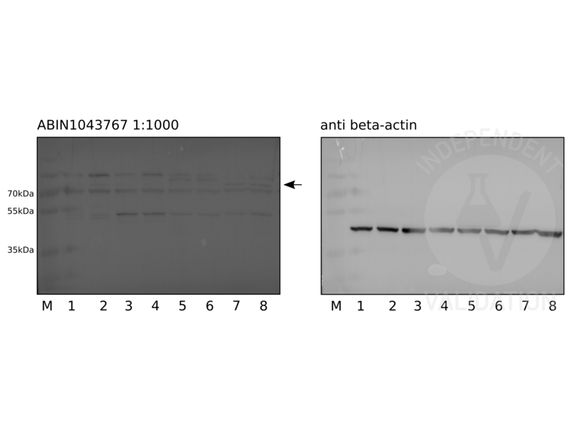
ABIN1043767 reveals a protein band of the expected MW and some weaker extraneous bands in cell lysates of human pluripotent stem cells and pancreatic progenitor cells.
- Primary Antibody
- ABIN1043767
- Secondary Antibody
- donkey anti-rabbit HRP-conjugated antibody (GE Healthcare, NA9310V)
- Full Protocol
- Grow HUES8 in mTeSR1 (STEMCELL Technologies, 85850) at 37°C and 5% O2, 5% CO2 in 2ml in a 6-well plate to 90% confluency.
- Harvest cells using TrypLE (ThermoFisher Scientific, 12604013) following the manufacturer´s instructions.
- Lyse 2x10⁶ cells in 30µl per well cold RIPA buffer (50mM Tris-HCl pH 8.0, 150mM NaCl, 0.1% SDS, 0.5% deoxycholate, 1% TritonX 100 in ddH2O) supplemented with 1mM PMSF and 1x protease inhibitor (Roche, 11836170001) for 30min on ice with 3x vortexing in 1.5ml microcentrifuge tubes.
- Centrifuge tubes at 10000xg for 8min at 4°C.
- Transfer supernatant to a new 1.5ml microcentrifuge tube and store at -80°C.
- Determine total protein content of the lysates using a Bradford assay (Bio-Rad, 500-0006).
- Denature 50µg of total protein for 5min at 95°C in 30µl 1x Laemmli SDS sample buffer and subsequently separate them on a denaturing 7.5% polyacrylamide gel (7.5% Acrylamide, 0.375M Trís-HCl (pH8.8), 0.1% SDS, 0.1% APS, 0.1% TEMED) for about 90min at 120V.
- Transfer proteins onto a PVDF membrane (Sigma Aldrich, IPVH00010) with transfer buffer (5.27g Tris, 2.93 g glycerine, 200 ml methanol, fill to 1l ddH2O) in a semidry western blotting system for 75min at 80mA/gel.
- Check transfer of the separated proteins by Ponceau S staining.
- Rinse membrane with water.
- Wash membrane for 5min with TBST (TBS, 0.02% Tween20).
- Block the membrane with 20ml blocking buffer (TBST, 5% milk) on a shaker for 1h at RT.
- Rinse membrane 3x with TBST.
- Wash membrane on a shaker 3x for 5min with TBST.
- Shrink-wrap and incubate membrane with primary
- rabbit anti-JAG1 antibody (antibodies-online, ABIN1043767, lot 28285) diluted 1:1000 and 1:10000 respectively in blocking buffer ON at 4°C or
- mouse anti-beta actin antibody (Sigma Aldrich, A1978) diluted 1:2000 in blocking buffer at RT for 1h.
- Wash membrane 3x for 5min with TBST.
- Incubate membrane with secondary
- donkey anti-rabbit HRP-conjugated antibody (GE Healthcare, NA9310V) diluted 1:5000 in TBST containing 1% milk for 1h at RT or
- donkey anti-mouse HRP-conjugated antibody (GE Healthcare, NA9340V) diluted 1:5000 in TBST containing 1% milk for 1h at RT.
- Wash membrane 3x for 5min with TBST.
- Reveal protein bands using ECL solution (ThermoScientific, 34076) on a Fusion SL (Vilber) chemiluminescence system. Exposure times for the 1:1000 and 1:10000 dilutions of ABIN1043767 were 30sec and 5min respectively.
- Experimental Notes
The bands marked by the arrow in the western blot image using ABIN1043767 at a 1:1000 dilution seem to be specific, as JAG1 showed highest transcriptomic levels at pancreatic progenitor stage (lanes 7 and 8), followed by pluripotent stem cell state (lane 2). Although the immunoblot with ABIN1043767 results in some unspecific bands, the expected band height fits nicely (75kDa). Higher MW bands might potentially represent precursor proteins detectable in all samples but pancreatic progenitor stage samples. In principle specific bands are also detectable with ABIN1043767 at a 1:10000 dilution, although exposition time was very long.
ABIN1043767 was also tested in IHC on 4-5µm FFPE sections of primary human kidney cancer tissue. Epitope retrieval was carried out using Tris-EDTA buffer at pH9.0 (Zytomed, ZUC029-500), EDTA at pH8.0 (Leica, RE7116), or citrate buffer at pH6.1 (Agilent, S169984-2) for 20min in a decloaking chamber. Antigen retrieval at pH8.0 looks ok, assuming a broad expression pattern. However, detection would be rather expected in fewer tubular cells. No staining was observed in glomeruli. Cytoplasmic staining in tubuli is revealed at higher magnification. Note that antigen retrieval influences the staining pattern. All in all, staining needs to be validated for example in protein ablation assays.
Validation #103702 (Western Blotting)
 Validation Images
![Western blot with ABIN1043686 on human pluripotent stem cells (1 and 2), definite endoderm cells (3 and 4), pancreatic endoderm cells (5 and 6), and pancreatic progenitors (7 and 8). ABIN1043686 was used at a 1:1000 (30sec exposure) or 1:10000 dilution (5min exposure). Beta-actin served as loading control.]() Western blot with ABIN1043686 on human pluripotent stem cells (1 and 2), definite endoderm cells (3 and 4), pancreatic endoderm cells (5 and 6), and pancreatic progenitors (7 and 8). ABIN1043686 was used at a 1:1000 (30sec exposure) or 1:10000 dilution (5min exposure). Beta-actin served as loading control.
Full Methods
Western blot with ABIN1043686 on human pluripotent stem cells (1 and 2), definite endoderm cells (3 and 4), pancreatic endoderm cells (5 and 6), and pancreatic progenitors (7 and 8). ABIN1043686 was used at a 1:1000 (30sec exposure) or 1:10000 dilution (5min exposure). Beta-actin served as loading control.
Full Methods -
- Format
- Liquid
- Concentration
- 1.1 mg/mL
- Buffer
-
Buffer: 0.02 M Potassium Phosphate, 0.15 M Sodium Chloride, pH 7.2
Stabilizer: None
Preservative: 0.01 % (w/v) Sodium Azide - Preservative
- Sodium azide
- Precaution of Use
- This product contains Sodium azide: a POISONOUS AND HAZARDOUS SUBSTANCE which should be handled by trained staff only.
- Storage
- 4 °C,-20 °C
- Storage Comment
- Store vial at -20° C prior to opening. Aliquot contents and freeze at -20° C or below for extended storage. Avoid cycles of freezing and thawing. Centrifuge product if not completely clear after standing at room temperature. This product is stable for several weeks at 4° C as an undiluted liquid. Dilute only prior to immediate use.
- Expiry Date
- 12 months
-
-
: "Notch-Jagged signalling can give rise to clusters of cells exhibiting a hybrid epithelial/mesenchymal phenotype." in: Journal of the Royal Society, Interface, Vol. 13, Issue 118, (2017) (PubMed).
: "Immunohistological localization of Notch receptors and their ligands Delta and Jagged in synovial tissues of rheumatoid arthritis." in: Journal of orthopaedic science : official journal of the Japanese Orthopaedic Association, Vol. 10, Issue 6, pp. 589-94, (2005) (PubMed).
-
: "Notch-Jagged signalling can give rise to clusters of cells exhibiting a hybrid epithelial/mesenchymal phenotype." in: Journal of the Royal Society, Interface, Vol. 13, Issue 118, (2017) (PubMed).
-
- Target
- JAG1 (Jagged 1 (JAG1))
- Alternative Name
- JAG1 (JAG1 Products)
- Background
- Background: Anti-Jagged 1 Antibody recognizes the Jagged 1 protein encoded by the JAG1 gene, that is the human homolog of the Drosophilia jagged protein. Human Jagged 1 is the ligand for multiple Notch receptors and mediation of Notch signaling. Jagged 1 signaling through Notch 1 has also been shown to play a role in hematopoiesis. Mutations that alter the Jagged 1 protein cause Alagille syndrome, deafness, congenital heart defects, and posterior embryotoxon. Anti-Jagged-1 Antibody is useful for researchers interested in Notch activation, Growth Factor activities, and Cardiovascular health.
- Gene ID
- 182, 4557679
- UniProt
- P78504
- Pathways
- Notch Signaling, Stem Cell Maintenance
-


